Applications & Publications
Protein arrays for your research
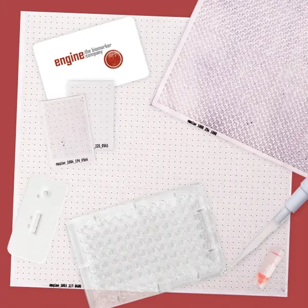
Protein arrays open up manifold possibilities as they can be used for analysing protein interactions with numerous types of molecules. You can determine auto-antibody profiles of complex sera, cross-reactivity of monoclonal antibodies, protein-protein interaction pairs, DNA/RNA-binding proteins, and much more.
For more than 20 years our protein arrays, also called high-density filters, have been used in biomarker research all over the world and for various indications. Whether you want to discover new biomarkers for diagnostics or therapy, or gain a deeper understanding of disease or metabolic pathways. Get an overview of your research area in our database of publications!
Research areas
Protein arrays accelerate your research from hypothesis to validation.
Diseases & pathways
- Apoptosis
- Autoimmune diseases
- Cancer
- Cardiovascular disease
- Corona virus
- Diabetes
- Food, microbiome, lifestyle
- Infection & vaccination
- Inflammation
- Kidney disease
- Neurobiology
- Signalling pathways
Target interactions
- Antibody profiling (e.g. IgM, IgG, IgA)
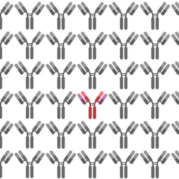
- Antibody specificity & off-targets
- DNA/RNA-binding
- Enzymatic modifications
- Immune response profiling
- Protein-protein interactions
- Small molecule profiling
Biomarker applications
- Companion diagnostics
- Diagnostics
- Drug discovery & therapeutics
- Monitoring
- Personalized medicine
- Risk stratification & prognostic
- Prediction & response
- Vaccination
Spotlighted publications
Case studies
Neurobiology
Validation of Plasma Proteomic Biomarkers Relating to Brain Amyloid Burden in the EMIF-Alzheimer’s Disease Multimodal Biomarker Discovery Cohort
Diagnosing Alzheimer’s disease at pre-symptomatic stages is the key to decelerating disease progression, but it is a costly and invasive process as it requires lumbar puncture. By screening patient plasma samples using protein arrays, mass spectrometry, and immunocaputre methods, the group could identify a 7‑protein biomarker panel which is predicted to reduce the screen failure rate by 20% .
Westwood, Sarah et al. “Validation of Plasma Proteomic Biomarkers Relating to Brain Amyloid Burden in the EMIF-Alzheimer’s Disease Multimodal Biomarker Discovery Cohort.” Journal of Alzheimer’s disease : JAD vol. 74,1 (2020): 213–225. doi:10.3233/JAD-190434
Corona virus
Gene expression profiling of corona virus microarray datasets to identify crucial targets in COVID-19 patients
In order to gain a better insight into the mechanism behind SARS-CoV immune dysregulation associated with high mortality, large amounts of microarray data were gathered. With the help of this dataset of normal and severe COVID-19 patients, a protein-protein network could be constructed. Hub genes could be identified (e.g CAMP, ELANE, LTF), the dysregulation of which is associated with high mortality rates.
Ramesh, Priyanka et al. “Gene expression profiling of corona virus microarray datasets to identify crucial targets in COVID-19 patients.” Gene reportsvol. 22 (2021): 100980. doi:10.1016/j.genrep.2020.100980
Cancer
Combination of Autoantibody Signature with PSA Level Enables a Highly Accurate Blood-Based Differentiation of Prostate Cancer Patients from Patients with Benign Prostatic Hyperplasia
Using Hex1 high-density protein macroarrays, sera of patients with prostate cancer were screened for disease-specific autoantibodies. The applied method can distinguish prostate cancer patients from normal controls with an accuracy of 83.2%, making the identified biomarkers are thus an important diagnostic tool.
Leidinger, Petra et al. “Combination of Autoantibody Signature with PSA Level Enables a Highly Accurate Blood-Based Differentiation of Prostate Cancer Patients from Patients with Benign Prostatic Hyperplasia.” PloS one vol. 10,6 e0128235. 3 Jun. 2015, doi:10.1371/journal.pone.0128235
Signalling pathways
Identification of a high-affinity network of secretagogin-binding proteins involved in vesicle secretion
The cellular interaction network of the protein secretagogin, which is known for its support for hierarchical organizational principles in the mammalian brain, was expanded by nine proteins through protein array screenings. The interactions of the identified proteins were verified by means of a GST pulldown assay. The interacting proteins form a specific network that mediates secretion and vesicle transport. This suggests a regulating role for secretagogin in these processes.
Bauer, Mikael C et al. “Identification of a high-affinity network of secretagogin-binding proteins involved in vesicle secretion.” Molecular bioSystemsvol. 7,7 (2011): 2196-204. doi:10.1039/c0mb00349b
Autoimmune disease
Immunogenicity of autoantigens
This paper examined which structural or biological features of certain auto-antigens and tumor-associated antigens trigger an immune response. In-silico methods were used to analyze large structural databases of previously identified antigens, identified with the help of various human protein array screenings. The authors state that proteins that are evolutionarily conserved, have certain sequence motifs or are part of cellular structures are more likely to trigger auto-immunogenic reactions.
Backes, Christina et al. “Immunogenicity of autoantigens.” BMC genomics vol. 12 340. 4 Jul. 2011, doi:10.1186/1471–2164-12–340
Free Brainstorming Session
Get in contact with our experts – We are happy to answer any question!
Protein Array Research Database
Science is based on sharing knowledge and building on the efforts of others. With this publication database, we would like to honour the valuable work of scientist – and support you in your research. We will be pleased if this provides you with inspiration, helpful information or even makes you aware of possible collaborations.
What’s inside?
- Protein array related publications, dealing with specific diseases such as spondyloarthritis
- Studies on overall indications such as autoimmunity
- Reviews and publications focussing arrays from the technical, methodological perspective
- Exciting papers with peptide arrays or other protein arrays
How to use the database
Search by full-text search or categories and keywords to find hits from your desired research field.
We are happy to include new publications! Feel free to contact us, if you have any study for our database. Please let us also know if something is not working :-).
Please note that the table is only shown in desktop version.
Simply enter your search term, e.g.:
- Autoimmune Diseases
- Cancer
- Biomarker Discovery
- Signalling Pathways
- Methods/Technology
Category | Keywords | Title | Author | Abstract | Journal | Year | Link |
---|---|---|---|---|---|---|---|
Pathogens | Chlamydophila | Gene expression profiling of corona virus microarray datasets to identify crucial targets in COVID-19 patients | P. Ramesh, S. Veerappapillai, R. Karuppasamy | The current outbreak of coronavirus disease (COVID-19) has been affecting millions of people and has caused devastating mortality worldwide. Moreover, it is to be noted that cytokine storm has become an important cause for the rising mortality. However, the efforts for the development of drugs, vaccines and treatment has also been intervened due to poor understanding of host’s defense mechanism and also due to the development of cytokine storm against this viral infection. Thus, a deeper understanding of the mechanism behind the immune dysregulation and cytokine storm development might give us clues for the clinical management of the severe cases. Hence, we have implemented differential gene expression analysis together with protein-protein interaction and Gene Ontology (GO) studies with the help of Severe Acute respiratory syndrome coronavirus (SARS-CoV) data sets such as GSE1739 and GSE33267 to give us more knowledge on the host immune response for the pathogenic coronavirus which in turn reduces the mortality. A total of 79 differentially-expressed genes (DEGs) were identified in our data set using the filters such as P‑value and log2 fold change values of less than 0.05 and 1.5 respectively. Further, network analysis and GO studies showed that differential expression of two hub genes namely ELANE and LTF which could induce higher levels of pro-inflammatory cytokines in the lungs. We are certain that differential expression of ELANE and LTF results in an excessive inflammatory reaction known as the cytokine storm and ultimately leading to death. Therefore, targeting these key drivers of cytokine storm genes appears to be the potential therapeutic targets for combating the Severe Acute respiratory syndrome coronavirus — 2 (SARS-CoV‑2) infection ultimately resulting in reduced mortality. Indeed, this predictive view may open new insights for designing an immune intervention for COVID-19 in the near future resulting in the mitigation of mortality rate. | Gene Reports | 2021 | https://doi.org/10.1016/j.genrep.2020.100980 |
Neurobiology | Alzheimer’s Disease, Biomarker Discovery | Validation of Plasma Proteomic Biomarkers Relating to Brain Amyloid Burden in the EMIF-Alzheimer’s Disease Multimodal Biomarker Discovery Cohort | S. Westwood, A. L. Baird, S. N. Anand, A. J. Nevado-Holgado, A. Kormilitzin, L. Shi , Abdul Hye, N. J. Ashton, A. R. Morgan, I. Bos, S. J. B. Vos, S. Baker, N. J. Buckley, M. Ten Kate, P. Scheltens, C. E. Teunissen, R. Vandenberghe, S. Gabel, K. Meersmans, S. Engelborghs, E. E. De Roeck, K. Sleegers, G. B. Frisoni, O. Blin, J. C. Richardson, R. Bordet, J. L. Molinuevo, L. Rami, A. Wallin, P. Kettunen, M. Tsolaki, F. Verhey, A. Lléo , I. Sala, J. Popp , G. Peyratout, P. Martinez-Lage, M. Tainta, P. Johannsen, Y. Freund-Levi, L. Frölich, V. Dobricic, C. Legido-Quigley, L. Bertram, F. Barkhof, H. Zetterberg, B. P. Morgan, J. Streffer, P. Jelle Visser, S. Lovestone | We have previously investigated, discovered, and replicated plasma protein biomarkers for use to triage potential trials participants for PET or cerebrospinal fluid measures of Alzheimer’s disease (AD) pathology. This study sought to undertake validation of these candidate plasma biomarkers in a large, multi-center sample collection. Targeted plasma analyses of 34 proteins with prior evidence for prediction of in vivo pathology were conducted in up to 1,000 samples from cognitively healthy elderly individuals, people with mild cognitive impairment, and in patients with AD-type dementia, selected from the EMIF-AD catalogue. Proteins were measured using Luminex xMAP, ELISA, and Meso Scale Discovery assays. Seven proteins replicated in their ability to predict in vivo amyloid pathology. These proteins form a biomarker panel that, along with age, could significantly discriminate between individuals with high and low amyloid pathology with an area under the curve of 0.74. The performance of this biomarker panel remained consistent when tested in apolipoprotein E ɛ4 non-carrier individuals only. This blood-based panel is biologically relevant, measurable using practical immunocapture arrays, and could significantly reduce the cost incurred to clinical trials through screen failure. | Journal of Alzheimer’s Disease | 2020 | https://doi.org/10.3233/jad-190434 |
Pathogens, Neurobiology | Gonorrhea, Exocytosis, Schizophrenia, Antigen Identification | Crossreactivity of an Antiserum Directed to the Gram-Negative Bacterium Neisseria gonorrhoeae with the SNARE-Complex Protein Snap23 Correlates to Impaired Exocytosis in SH-SY5Y Cells | A. Almamy, C. Schwerk, H. Schroten, H. Ishikawa, A. R. Asif, B. Reuss | Early maternal infections with Neisseria gonorrhoeae (NG) correlate to an increased lifetime schizophrenia risk for the offspring, which might be due to an immune-mediated mechanism. Here, we investigated the interactions of polyclonal antisera to NG (α‑NG) with a first trimester prenatal brain multiprotein array, revealing among others the SNARE-complex protein Snap23 as a target antigen for α‑NG. This interaction was confirmed by Western blot analysis with a recombinant Snap23 protein, whereas the closely related Snap25 failed to interact with α‑NG. Furthermore, a polyclonal antiserum to the closely related bacterium Neisseria meningitidis (α‑NM) failed to interact with both proteins. Functionally, in SH-SY5Y cells, α‑NG pretreatment interfered with both insulin-induced vesicle recycling, as revealed by uptake of the fluorescent endocytosis marker FM1-43, and insulin-dependent membrane translocation of the glucose transporter GluT4. Similar effects could be observed for an antiserum raised directly to Snap23, whereas a serum to Snap25 failed to do so. In conclusion, Snap23 seems to be a possible immune target for antigonococcal antibodies, the interactions of which seem at least in vitro to interfere with vesicle-associated exocytosis. Whether these changes contribute to the correlation between maternal gonococcal infections and psychosis in vivo remains still to be clarified. | Journal of Molecular Neuroscience | 2017 | https://doi.org/10.1007/s12031-017‑0920‑2 |
Cancer | Prostate Cancer, Diagnostics, Biomarker Discovery, Prostatic Hyperplasia | Combination of Autoantibody Signature with PSA Level Enables a Highly Accurate Blood- Based Differentiation of Prostate Cancer Patients from Patients with Benign Prostatic Hyperplasia | P. Leidinger, A. Keller, L. Milchram, C. Harz, M. Hart, A. Werth, H.-P. Lenhof, A. Weinhäusel, B. Keck, B. Wullich, N. Ludwig , E. Meese | Although an increased level of the prostate-specific antigen can be an indication for prostate cancer, other reasons often lead to a high rate of false positive results. Therefore, an additional serological screening of autoantibodies in patients’ sera could improve the detection of prostate cancer. We performed protein macroarray screening with sera from 49 prostate cancer patients, 70 patients with benign prostatic hyperplasia and 28 healthy controls and compared the autoimmune response in those groups. We were able to distinguish prostate cancer patients from normal controls with an accuracy of 83.2%, patients with benign prostatic hyperplasia from normal controls with an accuracy of 86.0%and prostate cancer patients from patients with benign prostatic hyperplasia with an accuracy of 70.3%. Combining seroreactivity pattern with a PSA level of higher than 4.0 ng/ml this classification could be improved to an accuracy of 84.1%. For selected proteins we were able to confirm the differential expression by using luminex on 84 samples. We provide a minimally invasive serological method to reduce false positive results in detection of prostate cancer and according to PSA screening to distinguish men with prostate cancer from men with benign prostatic hyperplasia. | PLOS One | 2015 | https://doi.org/10.1371/journal.pone.0128235 |
Cancer | PTM Identification, Monoclonal Gammopathies Of Undetermined Significance (MGUS), Multiple Myelomas (MM), Waldenstrom’s Macroglobulinemias (WM), Paraproteins | Sumoylated HSP90 is a dominantly inherited plasma cell dyscrasias risk factor | K.-D. Preuss, M. Pfreundschuh, N. Fadle, E. Regitz, B. Kubuschok | Posttranslationally modified proteins serve as autoimmunogenic targets in a wide spectrum of autoimmune diseases. Here, we identified a posttranslationally modified paraprotein target (paratargs) in monoclonal gammopathies of undetermined significance (MGUS), multiple myelomas (MM), and Waldenstrom’s macroglobulinemias (WM) using protein macroarrays that were sumoylated and screened for reactivity with paraproteins from MGUS, MM, and WM patients. We found that paraproteins from a proportion of European, African-American, and Japanese patients specifically reacted with the sumoylated heat-shock protein 90 β isoform‑α (HSP90-SUMO1, where SUMO indicates small ubiquitin-like modifier), while no reactivity with HSP90-SUMO1 was detected in over 800 controls. HSP90-SUMO1 was present in blood cells from all patients with HSP90-SUMO1-binding paraproteins. We determined that the HSP90-SUMO1 carrier state is autosomaldominantly inherited and caused by the inability of SUMO peptidase sentrin/SUMO-specific protease 2 (SENP2) to desumoylate HSP90-SUMO1. HSP90-SUMO1 was detected in a small percentage of healthy individuals from all backgrounds; however, only MGUS, MM, and WM patients who were HSP90-SUMO1 carriers produced HSP90-SUMO1-specific paraproteins, suggesting that sumoylated HSP90 promotes pathogenesis of these diseases through chronic antigenic stimulation. This study demonstrates that harboring HSP90-SUMO1 identifies healthy individuals at risk for plasma cell dyscrasias and that dominant inheritance of posttranslationally modified autoantigenic paratargs is one of the strongest molecular defined risk factors for MGUS, MM, and WM. | Journal of Clinical Investigation | 2015 | https://doi.org/10.1172/jci76802 |
Pathogens | Tuberculosis, NTM, Autoantibodies, Diagnostics, Mycobacterial Infection, Extrapulmonary | Autoantibodies binding to ubiquitin-fold modifier-conjugating enzyme 1 (Ufc1) and pleckstrin homology domain containing, family G (with RhoGef domain) member 2 (Plekhg2) are associated with mycobacterial infections | A. Tellermann, T. Witte, C. Lansche, M. Stoll, R. E. Schmidt, N. T. Baerlecken | The diagnosis of extrapulmonary tuberculous infections and nontuberculous mycobacterial (NTM) infections is difficult because the symptoms are nonspecific and suitable specimens for bacterial culture are often not available. Recent publications reported the existence of autoantibodies in tuberculous infections. We screened for specific autoantibodies in mycobacterial infections | HIV Medicine | 2015 | https://doi.org/10.1111/hiv.12194 |
Pathogens | Human Cytomegalovirus, Tumourigenesis, RNA Processing, Viral Gene Regulation | Identification of cellular proteins that interact with human cytomegalovirus immediate-early protein 1 by protein array assay | F. Puerta Martínez, Q. Tang | Human cytomegalovirus (HCMV) gene expression during infection is characterized as a sequential process including immediate-early (IE), early (E), and late (L)-stage gene expression. The most abundantly expressed gene at the IE stage of infection is the major IE (MIE) gene that produces IE1 and IE2. IE1 has been the focus of study because it is an important protein, not only for viral gene expression but also for viral replication. It is believed that IE1 plays important roles in viral gene regulation by interacting with cellular proteins. In the current study, we performed protein array assays and identified 83 cellular proteins that interact with IE1. Among them, seven are RNA-binding proteins that are important in RNA processing; more than half are nuclear proteins that are involved in gene regulations. Tumorigenesis-related proteins are also found to interact with IE1, implying that the role of IE1 in tumorigenesis might need to be reevaluated. Unexpectedly, cytoplasmic proteins, such as Golgi autoantigen and GGA1 (both related to the Golgi trafficking protein), are also found to be associated with IE1. We also employed a coimmunoprecipitation assay to test the interactions of IE1 and some of the proteins identified in the protein array assays and confirmed that the results from the protein array assays are reliable. Many of the proteins identified by the protein array assay have not been previously reported. Therefore, the functions of the IE1-protein interactions need to be further explored in the future. | Viruses | 2014 | http://doi.org/10.3390/v6010089 |
Signalling Pathways | Processing Bodies (P‑Bodies), mRNA Stability, Complex Formation, mRNA Homeostasis | LMKB/MARF1 localizes to mRNA processing bodies, interacts with Ge‑1, and regulates IFI44L gene expression. | D. B. Bloch, P. Li, E. G. Bloch, D. F. Berenson, R. L. Galdos, P. Arora, R. Malhotra, C. Wu, W. Yang | The mRNA processing body (P‑body) is a cellular structure that regulates the stability of cytoplasmic mRNA. MARF1 is a murine oocyte RNA-binding protein that is associated with maintenance of mRNA homeostasis and genomic stability. In this study, autoantibodies were used to identify Limkain B (LMKB), the human orthologue of MARF1, as a P‑body component. Indirect immunofluorescence demonstrated that Ge‑1 (a central component of the mammalian core-decapping complex) co-localized with LMKB in P‑bodies. Two-hybrid and co-immunoprecipitation assays were used to demonstrate interaction between Ge‑1 and LMKB. The C‑terminal 120 amino acids of LMKB mediated interaction with Ge‑1 and the N‑terminal 1094 amino acids of Ge‑1 were required for interaction with LMKB. LMKB is the first protein identified to date that interacts with this portion of Ge‑1. LMKB was expressed in human B and T lymphocyte cell lines; depletion of LMKB increased expression of IFI44L, a gene that has been implicated in the cellular response to Type I interferons. The interaction between LMKB/MARF1, a protein that contains RNA-binding domains, and Ge‑1, which interacts with core-decapping proteins, suggests that LMKB has a role in the regulation of mRNA stability. LMKB appears to have different functions in different cell types: maintenance of genomic stability in developing oocytes and possible dampening of the inflammatory response in B and T cells. | PLOS One | 2014 | https://doi.org/10.1371/journal.pone.0094784 |
Arthritis, Autoimmune Diseases | Spondyloarthritis (SA), Biomarker Discovery, Diagnostics, Autoantibodies | Autoantibodies against CD74 in spondyloarthritis | N. T. Baerlecken, S. Nothdorft, G. H. Stummvoll, J. Sieper, M. Rudwaleit, S. Reuter, T. Matthias, R. E. Schmidt, T. Witte | Spondyloarthritis (SpA) is a common debilitating in- flammatory disorder with a frequency of 1–2% in the European popula- tion. Establishing diagnosis however is oſten difficult, since abnormali- ties in conventional X‑ray develop with a latency of several years and only HLA-B27 has been established as a laboratory marker. With the exception of MRI, there are no sufficient tools for the early diagnosis of SpA. | Annals of the Rheumatic Diseases | 2013 | https://doi.org/10.1136/annrheumdis-2012–202208 |
Arthritis, Autoimmune Diseases | Autoantibodies, Biomarkers and Complex Regional Pain Syndrome | IgG Autoantibodies Against p29ING4 In Early-Stage Type I Complex Regional Pain Syndrome (CRPS) and In Other Inflammatory Diseases Involving The Joints | N. T. Baerlecken, T. Witte, R. E. Schmidt, C. Lansche, M- Bernateck, N. Pursche | Complex regional pain syndrome (CRPS, a.k.a. RSD) usually occurs after limb injury, especially fractures of the distal radius and elective hand surgery. Recent research has shown that some patients respond to treatment with immunglobulins supporting an autoimmune pathogenesis of CRPS. There are, however, no diagnostic markers of CRPS. | ACR/ARHP Annual Meeting | 2013 | https://acrabstracts.org/abstract/igg-autoantibodies-against-p29ing4-in-early-stage-type-i-complex-regional-pain-syndrome-crps-and-in-other-inflammatory-diseases-involving-the-joints/ |
Autoimmune Diseases | Adult-Onset Still’S Disease, Autoantibodies, Biomarker Discovery | Antibodies Against Drp‑4 and Macropain Subunit C2 As a Potential Marker OF AOSD | N. T. Baerlecken, N. Pursche, T. Witte, R. E. Schmidt, M. Hoepfner, F. Moosig, W. Gross, E. Feist, D. Foell | Making the diagnosis of adult-onset Still’s disease (AOSD) is mainly based on the exclusion of inflammatory, infectious and malignant diseases. There are no specific clinical or laboratory findings for AOSD. Therefore, we aimed to identify new autoantibodies as diagnostic tools for AOSD. | ACR/ARHP Annual Meeting | 2013 | https://acrabstracts.org/abstract/antibodies-against-drp-4-and-macropain-subunit-c2-as-a-potential-marker-of-aosd/ |
Neurobiology | Myasthenia Gravis, Nicotinic Acetylcholine Receptor, Autoantibodies, Myasthenia Gravis, Diagnostics, Biomarker Discovery, Autoantigens | Myasthenia gravis: analysis of serum autoantibody reactivities to 1827 potential human autoantigens by protein macroarrays | A. Becker, N. Ludwig, A. Keller, B. Tackenberg, C. Eienbröker, W. H. Oertel, K. Fassbender, E. Meese, K. Ruprecht | BACKGROUND: Myasthenia gravis is a disorder of neuromuscular transmission associated with autoantibodies against the nicotinic acetylcholine receptor. We have previously developed a customized protein macroarray comprising 1827 potential human autoantigens, which permitted to discriminate sera of patients with different cancers from sera of healthy controls, but has not yet been evaluated in antibody-mediated autoimmune diseases To determine whether autoantibody signatures obtained by protein macroarray separate sera of patients with myasthenia gravis from healthy controls. | PLOS One | 2013 | http://doi.org/10.1371/journal.pone.0058095 |
Arthritis, Autoimmune Diseases | Biomarker Discovery, Polymyalgia Rheumatica, Giant Cell Arteritis | Association of ferritin autoantibodies with giant cell arteritis/polymyalgia rheumatica | N. T. Baerlecken, A. Linnemann, W. L. Gross, F. Moosig, T. R. Vazquez-Rodriguez, M. A. Gonzalez-Gay, J. Martin, I. Kötter, J. C. Henes, I. Melchers, P. Vaith, R. E. Schmidt, T. Witte | Objectives Polymyalgia rheumatica (PMR) and giant cell arteritis (GCA) are relatively common inflammatory disorders. Establishing the diagnosis however may be difficult, since so far no specific biomarkers of the disorders are available. Methods: As a screening procedure, the authors used protein arrays for the detection of new autoantigens in GCA and PMR. The results of the protein array were confirmed by different ELISAs detecting IgG antibodies against the human ferritin heavy chain, N‑terminal 27 amino acids of the human ferritin heavy chain or the homologous peptide of Staphylococcus epidermidis. Sera of patients with only GCA (n=64), only PMR (n=47) and both PMR and GCA (n=31) were used. Results: In the ELISA using the human ferritin peptide, the sensitivity of IgG antibodies against ferritin was 92% in 36 GCA and/or PMR patients before initiation of treatment, 22/32 (69%) in patients with disease flares and 64/117 (55%) in the total cohort including treated and inactive patients. In controls, the false positive rate was 11/38 (29%) in systemic lupus erythematosus, 1/36 (3%) in rheumatoid arthritis, 0/31 (0%) in late onset rheumatoid arthritis, 3/46 (6.5%) in B‑non-Hodgkin’s lymphoma and 1/100 (1%) in blood donors. In the ELISA using the ferritin peptide of S epidermidis, 89% of 27 patients with untreated GCA and PMR were positive. Conclusion: Antibodies against the ferritin peptide were present in up to 92% of untreated, active GCA and PMR patients. They can be useful as a diagnostic marker of PMR and GCA. | Annals of the Rheumatic Diseases | 2012 | https://doi.org/10.1136/annrheumdis-2011–200413 |
Cancer | Biomarker Discovery, Ovarian Cancer, Autoantibodies, Diagnostics, p53 | Epitope presentation is an important determinant of the utility of antigens identified from protein arrays in the development of autoantibody diagnostic assays | M. A. Murphy, D. J. O’Connell, S. L. O’Kane, J. K. O’Brien, S. O’Toole, C. Martin, O. Sheils, J. J. O’Leary, D. J. Cahill | Autoantibodies represent an attractive biomarker for diagnostic assays principally due to the stability of immunoglobulin in patient serum facilitating measurement with conventional assays. Immune responses to tumorigenesis may facilitate detection of ovarian cancer in the early stages of the disease with identification of a panel of tumour specific autoantibodies. Despite the reporting of many tumour associated autoantibodies using arrays of tumour antigens, this has not led to the advance in diagnostic capability as rapidly as was initially expected. Here we examine the potential diagnostic utility of candidate autoantibody biomarkers identified via screening of serum samples on a high content human protein array from a unique cohort of early stage and late stage ovarian cancer patients. We analyse the performance of autoantibodies to the tumour suppressor protein p53 and the novel autoantigens alpha adducin and endosulfine alpha identified in our array screen. Each antigen has different performance characteristics using conventional ELISA format and Western blot immunoassay. The high attrition rate of promising autoantigens identified by array screening can in part be explained by the presentation of the epitope of the antigen in the subsequent method of validation and this study provides directions on maximising the potential of candidate biomarkers. This article is part of a Special Issue entitled: Translational Proteomics. | Journal of Proteomics | 2012 | http://doi.org/10.1016/j.jprot.2012.02.031 |
Neurobiology | Schizophrenia, Proteostasis, Depression, Biomarker Discovery, Genetic Association Study | Proteomic, genomic and translational approaches identify CRMP1 for a role in schizophrenia and its underlying traits | V. Bader, L. Tomppo, S. V. Trossbach, N. J. Bradshaw, I. Prikulis, S. R. Leliveld, C.-Y. Lin, K. Ishizuka, A. Sawa, A. Ramos, I. Rosa, Á. García, J. R. Requena, M. Hipolito, N. Rai, E. Nwulia, U. Henning, S. Ferrea, C. Luckhaus, J. Ekelund, J. Veijola, M.-R. Järvelin, W. Hennah, C. Korth | Schizophrenia is a chronic illness of heterogenous biological origin. We hypothesized that, similar to chronic progressive brain conditions, persistent functional disturbances of neurons would result in disturbed proteostasis in the brains of schizophrenia patients, leading to increased abundance of specific misfolded, insoluble proteins. Identification of such proteins would facilitate the elucidation of molecular processes underlying these devastating conditions. We therefore generated antibodies against pooled insoluble proteome of post-mortem brains from schizophrenia patients in order to identify unique, disease-specific epitopes. We successfully identified such an epitope to be present on collapsin-response mediator protein 1 (CRMP1) in biochemically purified, insoluble brain fractions. A genetic association analysis for the CRMP1 gene in a large Finnish population cohort (n = 4651) corroborated the association of physical and social anhedonia with the CRMP1 locus in a DISC1 (Disrupted-in-schizophrenia 1)-dependent manner. Physical and social anhedonia are heritable traits, present as chronic, negative symptoms of schizophrenia and severe major depression, thus constituting serious vulnerability factors for mental disease. Strikingly, lymphoblastoid cell lines derived from schizophrenia patients mirrored aberrant CRMP1 immunoreactivity by showing an increase of CRMP1 expression, suggesting its potential role as a blood-based diagnostic marker. CRMP1 is a novel candidate protein for schizophrenia traits at the intersection of the reelin and DISC1 pathways that directly and functionally interacts with DISC1. We demonstrate the impact of an interdisciplinary approach where the identification of a disease-associated epitope in post-mortem brains, powered by a genetic association study, is rapidly translated into a potential blood-based diagnostic marker. | Human Molecular Genetics | 2012 | https://www.ncbi.nlm.nih.gov/pmc/articles/PMC3529585/ |
Autoimmune Diseases | Autoantibodies, Computational Biology, Mechanism Discovery | Immunogenicity of autoantigens | C. Backes, N. Ludwig, P. Leidinger, C.n Harz, J. Hoffmann, A. Keller, E. Meese, H.-P. Lenhof | Autoantibodies against self-antigens have been associated not only with autoimmune diseases, but also with cancer and are even found in healthy individuals. The mechanism causing the autoantibody response remains elusive for the majority of the immunogenic antigens. To deepen the understanding of autoantibody responses, we ask whether natural-occurring, autoimmunity-associated and tumor-associated antigens have structural or biological features related to the immune response. To this end, we have carried out the most comprehensive in-silicio study of different groups of autoantigens including large antigen sets identified by our groups combined with publicly available antigen sets. | BMC Genomics | 2011 | https://doi.org/10.1186/1471–2164-12–340 |
Cancer | Neuroblastoma, Wilms Tumor, Childhood Cancer, Diagnostics, Immunoreactivity, Antigen Identification, Immunoreactivity, Antigen Identification | Autoantibody Signature Differentiates Wilms Tumor Patients from Neuroblastoma Patients | J. Schmitt, A. Keller, N. Nourkami-Tutdibi, S. Heisel, N. Habel, P. Leidinger, N. Ludwig, M. Gessler, N. Graf, F. Berthold, H‑P. Lenhof, E. Meese | Several studies report autoantibody signatures in cancer. The majority of these studies analyzed adult tumors and compared the seroreactivity pattern of tumor patients with the pattern in healthy controls. Here, we compared the autoimmune response in patients with neuroblastoma and patients with Wilms tumor representing two different childhood tumors. We were able to differentiate untreated neuroblastoma patients from untreated Wilms tumor patients with an accuracy of 86.8%, a sensitivity of 87.0% and a specificity of 86.7%. The separation of treated neuroblastoma patients from treated Wilms tumor patients’ yielded comparable results with an accuracy of 83.8%. We furthermore identified the antigens that contribute most to the differentiation between both tumor types. The analysis of these antigens revealed that neuroblastoma was considerably more immunogenic than Wilms tumor. The reported antigens have not been found to be relevant for comparative analyses between other tumors and controls. In summary, neuroblastoma appears as a highly immunogenic tumor as demonstrated by the extended number of antigens that separate this tumor from Wilms tumor | PLOS One | 2011 | http://doi.org/10.1371/journal.pone.0028951 |
Methods/Technology | Motifs, Affinity-Proteomic, Cross-Species, Mass Spectrometry | Proteomic Analysis and Discovery Using Affinity Proteomics and Mass Spectrometry | N. Olsson, C. Wingren, M. Mattsson, P. James, D. O’ Connell, F. Nilsson, D. J. Cahill, C. A. K. Borrebaeck | Antibody-based microarrays are a rapidly evolving affinity-proteomic methodology that recently has shown great promise in clinical applications. The resolution of these proteomic analyses is, however, directly related to the number of data-points, i.e. antibodies, included on the array. Currently, this is a key bottleneck because of limited availability of numerous highly characterized antibodies. Here, we present a conceptually new method, denoted global proteome survey, opening up the possibility to probe any proteome in a species-independent manner while still using a limited set of antibodies. We use context-independent-motif-specific antibodies directed against short amino acid motifs, where each motif is present in up to a few hundred different proteins. First, the digested proteome is exposed to these antibodies, whereby motif-containing peptides are enriched, which then are detected and identified by mass spectrometry. In this study, we profiled extracts from human colon tissue, yeast cells lysate, and mouse liver tissue to demonstrate proof-of-concept. | Molecular & Cellular Proteomics | 2011 | http://doi.org/10.1074/mcp.M110.003962 |
Signalling Pathways | PTM Identification, Transcription Factor, Mass Spectrometry | A differential proteome screening system for post-translational modification–dependent transcription factor interactions | O. Pless, E. Kowenz-Leutz, G. Dittmar & A. Leutz | Post-translational modifications (PTMs) of transcription factors alter interactions with co-regulators and epigenetic modifiers. For example, members of the C/EBP transcription factor family are extensively methylated on arginine and lysine residues in short, conserved, modular domains, implying modification-dependent cofactor docking. Here we describe array peptide screening (APS), a systematic and differential approach to detect PTM-dependent interactions in the human proteome using chemically synthesized, biotinylated peptides coupled to fluorophore-labeled streptavidin. Peptides with and without a modified residue are applied in parallel to bacterial expression libraries in an arrayed format. Interactions are detected and quantified by laser scanning to reveal proteins that differentially bind to nonmodified or modified peptides. We have previously used this method to investigate the effect of arginine methylation of C/EBPβ peptides. The method enables determination of PTM-dependent transcription factor interactions, quantification of relative binding affinities and rapid protein classification, all independently of the transactivation potential of peptides or cellular abundance of interactors. The protocol provides a cost-effective alternative to mass spectrometric approaches and takes 3–4 d to complete. | Nature Protocols | 2011 | https://doi.org/10.1038/nprot.2011.303 |
Signalling Pathways | Membrane Fusion, Secretion, Secratogogin, Vesicle Trafficking | Identification of a high-affinity network of secretagogin-binding proteins involved in vesicle secretion | M. C. Bauer, D. J. O’Connell, M. Maj, L. Wagner, D. J. Cahill, S. Linse | Secretagogin is a hexa EF-hand Ca(2+)-binding protein expressed in neuroendocrine, pancreatic endocrine and retinal cells. The protein has been noted for its expression in specific neuronal subtypes in the support of hierarchical organizing principles in the mammalian brain. Secretagogin has previously been found to interact with SNAP25 involved in Ca(2+)-induced exocytosis. Here, the cellular interaction network of secretagogin has been expanded with nine proteins: SNAP-23, DOC2alpha, ARFGAP2, rootletin, KIF5B, β‑tubulin, DDAH‑2, ATP-synthase and myeloid leukemia factor 2, based on screening of a high content protein array and validation and quantification of binding with surface plasmon resonance and GST pulldown assays. All targets have association rate constants in the range 10(4)-10(6) M(-1) s(-1), dissociation rate constants in the range 10(-3)-10(-5) s(-1) and equilibrium dissociation constants in the 100 pM to 10 nM range. The novel target SNAP23 is an essential component of the high affinity receptor for the general membrane fusion machinery and an important regulator of transport vesicle docking and fusion. Complementary roles in vesicle trafficking are known for ARFGAP2 and DOC2alpha in regulating fusion of vesicles to membranes, kinesin 5B and tubulin for transport of vesicles in the cell, while rootletin builds up the rootlet believed to function as a scaffold for vesicles. The identification of a discrete network of interacting proteins that mediate secretion and vesicle trafficking suggests a regulatory role for secretagogin in these processes. | Molecular BioSystems | 2011 | http://doi.org/10.1039/c0mb00349b |
Signalling Pathways | Redox Pathway, Platelet Glycoprotein, Thrombosis, Orphan Receptor | TNF receptor-associated factor 4 (TRAF4) is a novel binding partner of glycoprotein Ib and glycoprotein VI in human platelets | J. F. Arthur, Y. Shen, E. E. Gardiner, L. Coleman, D. Kenny, R. K. Andrews, M. C. Berndt | Background: Reactive oxygen species generation is one consequence of ligand engagement of platelet glycoprotein (GP) receptors GPIb-IX‑V and GPVI, which bind VWF/ collagen and initiate thrombosis at arterial shear; however, the precise molecular mechanism coupling redox pathway activa-tion to engagement of these receptors is unknown. Objective: The objective of this study was to identify novel binding partners for GPIb-IX‑V and GPVI that could provide a potential link between redox pathways and early platelet signaling events. Methods and Results: Using protein array analysis and affinity-binding assays, we demonstrated that the orphan TNF receptor-associated factor (TRAF) family mem-ber, TRAF4, selectively binds cytoplasmic sequences of GPIbb and GPVI. TRAF4, p47 phox [of the NADPH oxidase (Nox2) enzyme complex] and other redox relevant signaling proteins such as Hic‑5, co-immunoprecipitate with GPIb/GPVI from human platelet lysates whilst MBP-TRAF4 or MBP-p47 phox fusion proteins specifically pull-down GPIb/GPVI. GPIb-or GPVI-selective agonists induce phosphorylation of the TRAF4-associated proteins, Hic‑5 and Pyk2, with phosphor-ylation attenuated by Nox2 inhibition. Conclusion: These results describe the first direct association of TRAF4 with a receptor, and identify a novel binding partner for GPIb-IX‑V and GPVI, providing a potential link between these platelet receptors and downstream TRAF4/Nox2-dependent redox pathways. | Journal of Thrombosis and Haemostasis | 2011 | https://doi.org/10.1111/j.1538–7836.2010.04091.x |
Autoimmune Diseases, Neurobiology, Signalling Pathways | Glaucomna, Autoantigens, PXFG, Biomarker Discovery, Glaucoma, Neuroinflammation, Autoantibodies | Protein macroarray profiling of serum autoantibodies in pseudoexfoliation glaucoma | E. W. Dervan, H. Chen, S. Ling Ho, N. Brummel, J. Schmid, D. Toomey, M. Haralambova, E. Gould, D. M. Wallace, J. H. M. Prehn, C. J. O’Brien, D. Murphy | PURPOSE: Complex repertoires of IgG autoantibodies have been detected against ocular antigens in patients with glaucoma. The goal was to identify and characterize the IgG autoantibody repertoires in sera of patients with pseudoexfoliation glaucoma (PXFG) with protein macroarrays Complex repertoires of IgG autoantibodies have been detected against ocular antigens in patients with glaucoma. The goal was to identify and characterize the IgG autoantibody repertoires in sera of patients with pseudoexfoliation glaucoma (PXFG) with protein macroarrays. | Investigative Ophthalmology & Visual Science | 2010 | http://doi.org/10.1167/iovs.09–4898 |
Cancer | Machine Learning, Lung Cancer, Autoantibodies, Autoantigens, Diagnostics | Identification of lung cancer with high sensitivity and specificity by blood testing | P. Leidinger, A. Keller, S. Heisel, N. Ludwig, S. Rheinheimer, V. Klein, C. Andres, A. Staratschek-Jox, J. Wolf, E. Stoelben, B. Stephan, I. Stehle, J. Hamacher, H. Huwer, H.-P. Lenhof, E. Meese | Lung cancer is a very frequent and lethal tumor with an identifiable risk population. Cytological analysis and chest X‑ray failed to reduce mortality, and CT screenings are still controversially discussed. Recent studies provided first evidence for the potential usefulness of autoantigens as markers for lung cancer. | Respiratory Research | 2010 | http://doi.org/10.1186/1465–9921-11–18 |
Cancer, Methods/Technology | Biomarker Discovery, Monoclonal Antibodies, Immunostaining, Colorectal Cancer, Diagnostics, Antibody Profile | Human IgG antibody profiles differentiate between symptomatic patients with and without colorectal cancer | G. Kijanka, S. Hector, E. W. Kay, F. Murray, R. Cummins, D. Murphy, B. D. MacCraith, J. H. M. Prehn, D. Kenny | Objective: Patients with cancer have antibodies against tumour antigens. Characterising the antibody repertoire may provide insights into aberrant cellular mechanisms in cancer development, ultimately leading to novel diagnostic or therapeutic targets. The aim of this study was to characterise the antibody profiles in patients whose symptoms warranted colonoscopy, to see if there was a difference in patients with and without colorectal cancer. Methods: Patients were recruited from a colonoscopy clinic. Individual serum samples from 43 patients with colorectal cancer and 40 patients with no cancer on colonoscopy were profiled on a 37 830 clone recombinant human protein array. Antigen expression was evaluated by quantitative reverse transcription-PCR and by immunohistochemistry on tissue microarrays. Results: Using a sex- and age-matched training set, 18 antigens associated with cancer and 4 associated with the absence of cancer (p0.05) were identified and confirmed. To investigate the mechanisms triggering antibody responses to these antigens, antigen expression was examined in normal colorectal mucosa and colorectal carcinoma of the same patients. The identified antigens showed cellular accumulation (p53), aberrant cellular expression (high mobility group B1 (HMGB1)) and overexpression (tripartite motif-containing 28 (TRIM28), p53, HMGB1, transcription factor 3 (TCF3), longevity assurance gene homologue 5 (LASS5) and zinc finger protein 346 (ZNF346)) in colorectal cancer tissue compared with normal colorectal mucosa. Conclusions: It is demonstrated for the first time that screening high-density protein arrays identifies unique antibody profiles that discriminate between symptomatic patients with and without colorectal cancer. The differential expression of identified antigens suggests their involvement in aberrant cellular mechanisms in cancer. | Gut | 2010 | https://doi.org/10.1136/gut.2009.178574 |
Signalling Pathways | Transcription Factor, Extracellular Signalling, Methylation, Mass Spectrometry, Chromatin Remodelling, Differentiation, Histone Code, PTM Identification, Signalling, Epigenetics | Crosstalk between C/EBPbeta phosphorylation, arginine methylation, and SWI/SNF/Mediator implies an indexing transcription factor code | E. Kowenz-Leutz, O. Pless, G. Dittmar, M. Knoblich, A. Leutz | Cellular signalling cascades regulate the activity of transcription factors that convert extracellular information into gene regulation. C/EBPbeta is a ras/MAPkinase signal-sensitive transcription factor that regulates genes involved in metabolism, proliferation, differentiation, immunity, senescence, and tumourigenesis. The protein arginine methyltransferase 4 PRMT4/CARM1 interacts with C/EBPbeta and dimethylates a conserved arginine residue (R3) in the C/EBPbeta N‑terminal transactivation domain, as identified by mass spectrometry of cell-derived C/EBPbeta. Phosphorylation of the C/EBPbeta regulatory domain by ras/MAPkinase signalling abrogates the interaction between C/EBPbeta and PRMT4/CARM1. Differential proteomic screening, protein interaction studies, and mutational analysis revealed that methylation of R3 constraines interaction with SWI/SNF and Mediator complexes. Mutation of the R3 methylation site alters endogenous myeloid gene expression and adipogenic differentiation. Thus, phosphorylation of the transcription factor C/EBPbeta couples ras signalling to arginine methylation and regulates the interaction of C/EBPbeta with epigenetic gene regulatory protein complexes during cell differentiation. | The EMBO Journal | 2010 | http://doi.org/10.1038/emboj.2010.3 |
Methods/Technology | Soluble Proteins, Crystallization, Esprit Technology, Subcloning, Directed Evolution, Protein Structure, Protein Expression | ESPRIT: An automated, library-based method for mapping and soluble expression of protein domains from challenging targets | H. Yumerefendi, F. Tarendeau, P. J. Mas, D. J. Hart | Expression of sufficient quantities of soluble protein for structural biology and other applications is often a very difficult task, especially when multimilligram quantities are required. In order to improve yield, solubility or crystallisability of a protein, it is common to subclone shorter genetic constructs corresponding to single- or multi-domain fragments. However, it is not always clear where domain boundaries are located, especially when working on novel targets with little or no sequence similarity to other proteins. Several methods have been described employing aspects of directed evolution to the recombinant expression of challenging proteins. These combine the construction of a random library of genetic constructs of a target with a screening or selection process to identify solubly expressing protein fragments. Here we review several datasets from the ESPRIT (Expression of Soluble Proteins by Random Incremental Truncation) technology to provide a view on its capabilities. Firstly, we demonstrate how it functions using the well-characterised NF-??B p50 transcription factor as a model system. Secondly, application of ESPRIT to the challenging PB2 subunit of influenza polymerase has led to several novel atomic resolution structures; here we present an overview of the screening phase of that project. Thirdly, analysis of the human kinase TBK1 is presented to show how the ESPRIT technology rapidly addresses the compatibility of challenging targets with the Escherichia coli expression system. | Journal of Structural Biology | 2010 | http://doi.org/10.1016/j.jsb.2010.02.021 |
Neurobiology, Signalling Pathways | Calmodulin, Membrane Proteins, Protein-Protein Interaction, Amino Acid Motifs, Pathways, Calmodulin | Integrated protein array screening and high throughput validation of 70 novel neural calmodulin-binding proteins | D. J. O’Connell, M. C. Bauer, J. O’Brien, W. M. Johnson, C. A. Divizio, S. L. O’Kane, T. Berggård, A. Merino, K. S. Åkerfeldt, S. Linse, D. J. Cahill | Calmodulin is an essential regulator of intracellular processes in response to extracellular stimuli mediated by a rise in Ca2+ion concentration. To profile protein-protein interactions of calmodulin in human brain, We probed a high content human protein array with fluorophore-labeled calmodulin in the presence of Ca2+. This protein array contains 37,200 redundant proteins, incorporating over 10,000 unique human neural proteins from a human brain cDNA library. We designed a screen to find high affinity (KD 1 ?M) binding partners of calmodulin and identified 76 human proteins from all intracellular compartments of which 72 are novel. We measured the binding kinetics of 74 targets with calmodulin using a high throughput surface plasmon resonance assay. Most of the novel calmodulin-target complexes identified have low dissociation rates (koff 10?3s?1) and high affinity (KD 1 M), consistent with the design of the screen. Many of the identified proteins are known to assemble in neural tissue, forming assemblies such as the spectrin scaffold and the postsynaptic density. We developed a microarray of the identified target proteins with which we can characterize the biochemistry of calmodulin for all targets in parallel. Four novel targets were verified in neural cells by co-immunoprecipitation, and four were selected for exploration of the calmodulin-binding regions. Using synthetic peptides and isothermal titration calorimetry, calmodulin binding motifs were identified in the potassium voltage-gated channel Kv6.1 (residues 474–493), calmodulin kinase-like vesicle-associated protein (residues 302–316), EF-hand domain family member A2 (residues 202– 216), and phosphatidylinositol-4-phosphate 5‑kinase, type I,?(residues 400–415). | Molecular & Cellular Proteomics | 2010 | http://doi.org/10.1074/mcp.M900324-MCP200 |
Signalling Pathways | Pathways, Molecular Signalling, Protein-Protein Interaction, Small Interfering RNAs, NF-kappaB Pathway, Extracellular Signalling, Intercellular Messaging, Transcription Factor, Gene Regulation | Expanding the Substantial Interactome of NEMO Using Protein Microarrays | B. J. Fenner, M. Scannell, J. H. M. Prehn | Signal transduction by the NF-kappaB pathway is a key regulator of a host of cellular responses to extracellular and intracellular messages. The NEMO adaptor protein lies at the top of this pathway and serves as a molecular conduit, connecting signals transmitted from upstream sensors to the downstream NF-kappaB transcription factor and subsequent gene activation. The position of NEMO within this pathway makes it an attractive target from which to search for new proteins that link NF-kappaB signaling to additional pathways and upstream effectors. In this work, we have used protein microarrays to identify novel NEMO interactors. A total of 112 protein interactors were identified, with the most statistically significant hit being the canonical NEMO interactor IKKbeta, with IKKalpha also being identified. Of the novel interactors, more than 30% were kinases, while at least 25% were involved in signal transduction. Binding of NEMO to several interactors, including CALB1, CDK2, SAG, SENP2 and SYT1, was confirmed using GST pulldown assays and coimmunoprecipitation, validating the initial screening approach. Overexpression of CALB1, CDK2 and SAG was found to stimulate transcriptional activation by NF-kappaB, while SYT1 overexpression repressed TNFalpha-dependent NF-kappaB transcriptional activation in human embryonic kidney cells. Corresponding with this finding, RNA silencing of CDK2, SAG and SENP2 reduced NF-kappaB transcriptional activation, supporting a positive role for these proteins in the NF-kappaB pathway. The identification of a host of new NEMO interactors opens up new research opportunities to improve understanding of this essential cell signaling pathway. | PLOS One | 2010 | http://doi.org/10.1371/journal.pone.0008799 |
Signalling Pathways | A‑Integrins, Cell Biology, Peptide-Affinity Chromatography, Platelet, Protein-Protein Interaction | Protein interactions with the platelet integrin αIIb regulatory motif | M. Raab, H. Daxecker, R. J. Edwards, A. Treumann, D. Murphy, N. Moran | Integrins are transmembrane proteins regulating cellular shape, mobility and the cell cycle. A highly conserved signature motif in the cytoplasmic tail of the integrin alpha-subunit, KXGFFKR, plays a critical role in regulating integrin function. To date, six proteins have been identified that target this motif of the platelet-specific integrin alpha(IIb)beta(3). We employ peptide-affinity chromatography followed-up with LC-MS/MS analysis as well as protein chips to identify new potential regulators of integrin function in platelets and put them into their biological context using information from protein:protein interaction (PPI) databases. Totally, 44 platelet proteins bind with high affinity to an immobilized LAMWKVGFFKR-peptide. Of these, seven have been reported in the PPI literature as interactors with integrin alpha-subunits. 68 recombinant human proteins expressed on the protein chip specifically bind with high affinity to biotin-tagged alpha-integrin cytoplasmic peptides. Two of these proteins are also identified in the peptide-affinity experiments, one is also found in the PPI databases and a further one is present in the data to all three approaches. Finally, novel short linear interaction motifs are common to a number of proteins identified. | Proteomics | 2010 | https://doi.org/10.1002/pmic.200900621 |
Autoimmune Diseases | COPD, Respiratory Disease, Diagnostics, Antibody Profile, Respiratory Disease, Autoantibodies | Novel autoantigens immunogenic in COPD patients | P. Leidinger, A. Keller, S. Heisel, N. Ludwig, S. Rheinheimer, V. Klein, C. Andres, J. Hamacher, H. Huwer, B. Stephan, I. Stehle, H.-P. Lenhof, E. Meese | Chronic obstructive pulmonary disease (COPD) is a respiratory inflammatory condition with autoimmune features including IgG autoantibodies. In this study we analyze the complexity of the autoantibody response and reveal the nature of the antigens that are recognized by autoantibodies in COPD patients. RESULTS: By visual evaluation we detected 381 peptide clones that reacted with autoantibodies of COPD patients including 17 clones that reacted with more than 60% of the COPD sera and seven clones that reacted with more than 90% of the COPD sera. The comparison of COPD sera and controls by the automated image analysis system identified 212 peptide clones with informative AUC values. By in silico sequence analysis we found an enrichment of sequence motives previously associated with immunogenicity | Respiratory Research | 2009 | http://doi.org/10.1186/1465–9921-10–20 |
Cancer | Glioma, Machine Learning, Antibody Profile, Glioma, Autoantibodies, Diagnostics, Personalized Medicine | Improving Seroreactivity-Based Detection of Glioma | N. Ludwig, A. Keller, S. Heisel, P. Leidinger, V. Klein, S. Rheinheimer, C. U. Andres, B. Stephan, W.-I. Steudel, N. M. Graf, B. Burgeth, J. Weickert, H.-P. Lenhof, E. Meese | Seroreactivity profiling emerges as valuable technique for minimal invasive cancer detection. Recently, we provided first evidence for the applicability of serum profiling of glioma using a limited number of immunogenic antigens. Here, we screened 57 glioma and 60 healthy sera for autoantibodies against 1827 Escherichia coli expressed clones, including 509 in-frame peptide sequences. By a linear support vector machine approach, we calculated mean specificity, sensitivity, and accuracy of 100 repetitive classifications. We were able to differentiate glioma sera from sera of the healthy controls with a specificity of 90.28%, a sensitivity of 87.31% and an accuracy of 88.84%. We were also able to differentiate World Health Organization grade IV glioma sera from healthy sera with a specificity of 98.45%, a sensitivity of 80.93%, and an accuracy of 92.88%. To rank the antigens according to their information content, we computed the area under the receiver operator characteristic curve value for each clone. Altogether, we found 46 immunogenic clones including 16 in-frame clones that were informative for the classification of glioma sera versus healthy sera. For the separation of glioblastoma versus healthy sera, we found 91 informative clones including 26 in-frame clones. The best-suited in-frame clone for the classification glioma sera versus healthy sera corresponded to the vimentin gene (VIM) that was previously associated with glioma. In the future, autoantibody signatures in glioma not only may prove useful for diagnosis but also offer the prospect for a personalized immune-based therapy. | Neoplasia | 2009 | http://doi.org/10.1593/neo.91018 |
Cancer, Methods/Technology | Histopathology, Nuclear Mitotic Apparatus Protein 1, Spindle Apparatus, Mitosis, Centrosome, Lymphoma, Colorectal Cancer, Pre-Symptom, Autoantibodies | Defining the molecular target of an antibody derived from nuclear extract of Jurkat cells using protein arrays | G. Kijankaa, R. Barry, H. Chen, E. Gould, S. K. Seidlits, J. Schmida, M. Morgan, D. Y. Mason, J. Cordell, D. Murphy | Many diagnostic antibodies are generated by immunization with whole cells or cell extracts and are shown by screening on tissue sections to label specific cell populations. However, their target molecule then needs to be identified, and this can be technically demanding. Here we describe the use of protein arrays to define the targets of new or uncharacterized monoclonal antibodies. The technique involves screening protein arrays containing thousands of recombinant human proteins. An initial experiment showed that a well-characterized monoclonal antibody against nucleophosmin identified 22 clones on the array encoding this protein. Next, the antibody JJ166, for which the antigen had not yet been identified, was screened. This antibody was generated by immunizing with a nuclear extract of Jurkat cells and was detected in immunohistochemistry due to its distinctive nuclear staining of lymphoid tissue cells. However, its molecular target had remained unidentified using traditional approaches. A protein array screen rapidly identified the mitotic spindle-associated molecule NUMA1 (nuclear mitotic apparatus protein 1). To confirm this putative specificity, JJ166 was shown to react with COS‑1 cells transiently transfected with the complementary DNA for NUMA1. Furthermore, a commercially available antibody against NUMA1 showed nearly identical staining in immunohistochemistry on human tissue and cells. Overall, this method represents an effective and quick strategy for defining the protein targets of new or uncharacterized monoclonal antibodies identified as having diagnostic or other potential value on the basis of their immunostaining patterns. | Analytical Biochemistry | 2009 | http://doi.org/10.1016/j.ab.2009.08.039 |
Methods/Technology | Epitope Mapping, Cross Reactions, Commercial Antibodies, Quality Control, Antibody Specificity, Monoclonal, Polyclonal | Rapid characterization of binding specificity and cross-reactivity of antibodies using recombinant human protein arrays | G. Kijanka, S. IpCho, S. Baars, H. Chen, K. Hadley, A. Beveridge, E. Gould, D. Murphy | Antibodies are routinely used as research tools, in diagnostic assays and increasingly as therapeutics. Ideally, these applications require antibodies with high sensitivity and specificity; however, many commercially available antibodies are limited in their use as they cross-react with non-related proteins. Here we describe a novel method to characterize antibody specificity. Six commercially available monoclonal and polyclonal antibodies were screened on high-density protein arrays comprising of ~ 10,000 recombinant human proteins (Imagenes). Two of the six antibodies examined; anti-pICln and anti-GAPDH, bound exclusively to their target antigen and showed no cross-reactivity with non-related proteins. However, four of the antibodies, anti-HSP90, anti-HSA, anti-bFGF and anti-Ro52, showed strong cross-reactivity with other proteins on the array. Antibody–antigen interactions were readily confirmed using Western immunoblotting. In addition, the redundant nature of the protein array used, enabled us to define the epitopic region within HSP90 of the anti-HSP90 antibody, and identify possible shared epitopes in cross-reacting proteins. In conclusion, high-density protein array technology is a fast and effective means for determining the specificity of antibodies and can be used to further improve the accuracy of antibody applications. | Journal of Immunological Methods | 2009 | http://doi.org/10.1016/j.jim.2008.10.008 |
Signalling Pathways | Polyubiquitination, NF-kappaB Pathway, Gene Expression Regulation, Ubiquitin, Polyubiquitin, Protein Metabolism | Identification of polyubiquitin binding proteins involved in NF-kappaB signaling using protein arrays | B. J. Fenner, M. Scannell, J. H. M. Prehn | Attachment of ubiquitin to proteins represents a central mechanism for the regulation of protein metabolism and function. In the NF-kappaB pathway, binding of NEMO to polyubiquitinated substrates initiates the pathway in response to cellular stimuli. Other polyubiquitin binding proteins can antagonize this pathway by competing with NEMO for polyubiquitin. We have used protein arrays to identify polyubiquitin binding proteins that regulate NF-kappaB activity. Using polyubiquitin as bait, protein arrays were screened and polyubiquitin binders identified. Novel polyubiquitin binders AWP1, CALCOCO2, N4BP1, RIO3, TEX27, TTC3, UBFD1 and ZNF313 were identified using this approach, while known NF-kappaB regulators including NEMO, A20, ABIN‑1, ABIN‑2, optineurin and p62 were also identified. Overexpressed AWP1 and RIO3 repressed NF-kappaB activity in a manner similar to optineurin, while siRNAs directed against AWP1 and RIO3 also reduced NF-kappaB activity. TNFalpha-dependent degradation of IkappaBalpha was also suppressed by overexpression of AWP1 and RIO3, possibly due to the polyubiquitin binding activity of these proteins. Protein array screening using polyubiquitin enabled rapid identification of many known and novel polyubiquitin binding proteins and the identification of novel NF-kappaB regulators. | Biochimica et Biophysica Acta | 2009 | http://doi.org/10.1016/j.bbapap.2009.02.013 |
Cancer, Signalling Pathways | Breast Cancer, Tyrosine Kinase, BRK Signalling Pathway, Signal Transduction, KAP3A, Protein Array | Breast tumor kinase BRK requires kinesin‑2 subunit KAP3A in modulation of cell migration | K. E. Lukong, S. Richard | Breast tumor Kinase (BRK) also known as protein kinase 6 (PTK6) is a nonreceptor tyrosine kinase overexpressed in the majority of human breast tumors. Although some studies have implicated BRK in signalling, cell proliferation and migration, the precise intracellular role of BRK has not been fully elucidated. The RNA-binding protein Sam68, and adaptor proteins paxillin and STAT3 are the only BRK substrates that link BRK to signal transduction. To identify new BRK substrates, we screened high-density protein filter arrays by large-scale in vitro kinase assays using active recombinant BRK. We identified at least 4 BRK targets comprising the alpha-subunit of stimulatory guanine nucleotide binding protein (GNAS), FL139441, ??-tubulin and kinesin associated protein 3A (KAP3A) and validated them as BRK substrates using a secondary assay. Further characterization revealed that KAP3A is an in vivo substrate of BRK and associates with BRK in breast cancer cells. We show that BRK specifically phosphorylated tyrosine residues at the C‑terminus of KAP3A and induces delocalization of KAP3A from punctate nuclear localization to a diffuse nucleo-cytoplasmic pattern. Functionally, we demonstrate that KAP3A knockdown results in suppression of BRK-induced migration of breast cancer cells and show that the C‑terminal deletion mutant of KAP3A acts as a dominant negative in BRK-induced cell migration. Our findings therefore reveal new substrates of BRK and define KAP3A as a physiological substrate of BRK during cell migration. ?? 2007 Elsevier Inc. All rights reserved. Breast Cancer, tyrosine kinase, BRK signalling pathway | Cellular Signalling | 2008 | http://doi.org/10.1016/j.cellsig.2007.11.003 |
Cancer, Signalling Pathways | Cyclin-Dependent Kinases, Substrate Discovery, Cell Migration, Breast Cancer, Molecular Pathways, SIRT2, Microtubules, PTM Identification | The regulation of SIRT2 function by cyclin-dependent kinases affects cell motility | R. Pandithage, R. Lilischkis, K. Harting, A. Wolf, B. Jedamzik, J. Lüscher-Firzlaff, J. Vervoorts, E. Lasonder, E. Kremmer, B. Knöll, B. Lüscher | Cyclin-dependent kinases (Cdks) fulfill key functions in many cellular processes, including cell cycle progression and cytoskeletal dynamics. A limited number of Cdk substrates have been identified with few demonstrated to be regulated by Cdk-dependent phosphorylation. We identify on protein expression arrays novel cyclin E‑Cdk2 substrates, including SIRT2, a member of the Sirtuin family of NAD(+)-dependent deacetylases that targets alpha-tubulin. We define Ser-331 as the site phosphorylated by cyclin E‑Cdk2, cyclin A‑Cdk2, and p35-Cdk5 both in vitro and in cells. Importantly, phosphorylation at Ser-331 inhibits the catalytic activity of SIRT2. Gain- and loss-of-function studies demonstrate that SIRT2 interfered with cell adhesion and cell migration. In postmitotic hippocampal neurons, neurite outgrowth and growth cone collapse are inhibited by SIRT2. The effects provoked by SIRT2, but not those of a nonphosphorylatable mutant, are antagonized by Cdk-dependent phosphorylation. Collectively, our findings identify a posttranslational mechanism that controls SIRT2 function, and they provide evidence for a novel regulatory circuitry involving Cdks, SIRT2, and microtubules. | Journal of Cell Biology | 2008 | http://doi.org/10.1083/jcb.200707126 |
Signalling Pathways | Gene Regulation, Methylation, PTM Identification, Enhancer-Binding Protein | G9a-mediated Lysine Methylation Alters the Function of CCAAT/Enhancer-binding Protein‑β | O. Pless, E. Kowenz-Leutz, M. Knoblich, J. Lausen, M. Beyermann, M.J. Walsh, A. Leutz | The functional capacity of the transcriptional regulatory CCAAT/enhancer-binding protein-beta (C/EBPbeta) is governed by protein interactions and post-translational protein modifications. In a proteome-wide interaction screen, the histone-lysine N‑methyltransferase, H3 lysine 9‑specific 3 (G9a), was found to directly interact with the C/EBPbeta transactivation domain (TAD). Binding between G9a and C/EBPbeta was confirmed by glutathione S‑transferase pulldown and co-immunoprecipitation. Metabolic labeling showed that C/EBPbeta is post-translationally modified by methylation in vivo. A conserved lysine residue in the C/EBPbeta TAD served as a substrate for G9a-mediated methylation. G9a, but not a methyltransferase-defective G9a mutant, abrogated the transactivation potential of wild type C/EBPbeta. A C/EBPbeta TAD mutant that contained a lysine-to-alanine exchange was resistant to G9a-mediated inhibition. Moreover, the same mutation conferred super-activation of a chromatin-embedded, endogenous C/EBPbeta target gene. Our data identify C/EBPbeta as a direct substrate of G9a-mediated post-translational modification that alters the functional properties of C/EBPbeta during gene regulation. | Journal of Biological Chemistry | 2008 | https://doi.org/10.1074/jbc.m802132200 |
Autoimmune Diseases | mRNA Processing, Gene Silencing, Primary Biliary Cirrhosis, mRNA Processing Bodies, Stress Granule, mRNA Decay, Autoantibodies, Primary Biliary Cirrhosis | Probing the mRNA processing body using protein macroarrays and “autoantigenomics” | W.-H. Yang, D. B. Bloch | Messenger RNA processing bodies (P‑bodies) are cellular structures that have a direct role in mRNA degradation. P‑bodies have also been implicated in RNAi-mediated post-transcriptional gene silencing. Despite the important roles of P‑bodies in cellular biology, the constituents of P‑bodies and their organization have been only partially defined. Approximately 5% of patients with the autoimmune disease primary biliary cirrhosis have antibodies directed against these structures. Recent advances in protein macroarray technology permit the simultaneous screening of thousands of proteins for reactivity with autoantibodies. We used serum from patients with anti-P-body autoantibodies to screen a protein macroarray and identified 67 potential autoantigens. Immunoreactive proteins included four known P‑body components and three additional primary biliary cirrhosis autoantigens. Y‑box protein 1 (YB‑1), a 50-kDa RNA-binding protein that was not previously known to be a P‑body component, was recognized by serum from four of seven patients. YB‑1 colocalized with P‑body components DCP1a and Ge‑1. In cells subjected to arsenite-induced oxidative stress, YB‑1 localized to TIA-containing stress granules. Both YB‑1 and the previously identified P‑body component RAP55 translocated from P‑bodies to stress granules during oxidative stress. During recovery, however, the reappearance of YB‑1 in P‑bodies was delayed compared with that of RAP55, suggesting that YB‑1 and RAP55 may have different functions. This study demonstrates that the combination of human autoantibodies and protein macroarray technology provides a novel method for identifying and characterizing components of mRNA P‑bodies. | RNA | 2007 | https://pubmed.ncbi.nlm.nih.gov/17339575/ |
Autoimmune Diseases | Cardiomyopathy, Antibody Profile, Autoantibodies, Dilated Cardiomyopathy | Profiling humoral autoimmune repertoire of dilated cardiomyopathy (DCM) patients and development of a disease-associated protein chip | S. Horn, A. Lueking, D. Murphy, A. Staudt, C. Gutjahr, K. Schulte, A. König, M. Landsberger, H. Lehrach, S. B. Felix, D. J. Cahill | Dilated cardiomyopathy (DCM) is a myocardial disease characterized by progressive depression of myocardial contractile function and ventricular dilatation. Thirty percent of DCM patients belong to the inherited genetic form; the rest may be idiopathic, viral, autoimmune, or immune-mediated associated with a viral infection. Disturbances in humoral and cellular immunity have been described in cases of myocarditis and DCM. A number of autoantibodies against cardiac cell proteins have been identified in DCM. In this study, we have profiled the autoantibody repertoire of plasma from DCM patients against a human protein array consisting of 37,200 redundant, recombinant human proteins and performed qualitative and quantitative validation of these putative autoantigens on protein microarrays to identify novel putative DCM specific autoantigens. In addition to analyzing the whole IgG autoantibody repertoire, we have also analyzed the IgG3 antibody repertoire in the plasma samples to study the characteristics of IgG3 subclass antibodies. By combining screening of a protein expression library with protein microarray technology, we have detected 26 proteins identified by the IgG antibody repertoire and 6 proteins bound by the IgG3 subclass. Several of these autoantibodies found in plasma of DCM patients, such as the autoantibody against the Kv channel-interacting protein, are associated with heart failure. | Proteomics | 2006 | http://doi.org/10.1002/pmic.200401293 |
Autoimmune Diseases, Cancer, Pathogens | MALT, Gastritis, Tumour Microenvironment, Neoplasia, Lymphoma, Mucosa-Associated Lymphoid Tissue, Helicobacter Pylori | Influence of antigen on the development of MALT lymphoma | D. Lenze, E. Berg, R. Volkmer-Engert, A. A. Weiser, A. Greiner, C. Knörr-Wittmann, I. Anagnostopoulos, H. Stein, M. Hummel | Mucosa-associated lymphoid tissue (MALT) B‑cell lymphomas develop in the context of autoimmune or chronic inflammations like Helicobacter pylori-induced gastritis. Remission of most gastric MALT lymphomas after eradication of H pylori links tumor cell proliferation to antigen-induced inflammation and the need for antigenic contact. Furthermore, the tumor cells correspond to antigen-activated memory B cells. To investigate the reactivity of the tumor immunoglobulins we employed in vitro-generated antibodies identical to those produced by MALT lymphoma cells. The immunoglobulin rearrangements of 7 MALT lymphomas were amplified, cloned, and expressed as single-chain fragment variable (scFv) antibodies. Antigen specificity of these 7 scFvs was analyzed by immunohistochemical staining of various normal, reactive, and malignant human tissues. Also, an expression library comprising approximately 30,000 proteins from human fetal brains (protein filter) and a peptide library were screened. One scFv stained a subpopulation of tonsillar plasma cells in immunohistochemical studies. On protein filters this scFv recognized the plasma cell-related protein Ufc1. Peptide library screening identified 9 peptides as binding partners of an additional scFv. The majority of MALT lymphoma immunoglobulins studied, however, showed no reactivity against antigens, indicating that the tumor immunoglobulins do not play a significant role in stimulation and proliferation of the MALT lymphoma tumor cells. | Blood | 2006 | http://doi.org/10.1182/blood-2005–04-1722 |
Signalling Pathways | Regulatory Network, Protein Array, Chip, Interaction Network, Epitope Mapping, Protein-Protein Interaction | Identification of VCP/p97, Carboxyl Terminus of Hsp70-interacting Protein (CHIP), and Amphiphysin II Interaction Partners Using Membrane-based Human Proteome Arrays | G. Grelle, S. Kostka, A. Otto, B. Kersten, K. F. Genser, E.-C. Müller, S. Wälter, A. Böddrich, U. Stelzl, C. Hänig, R. Volkmer-Engert, C. Landgraf, S. Alberti, J. Höhfeld, M. Strödicke, E. E. Wanker | Proteins mediate their biological function through interactions with other proteins. Therefore, the systematic identification and characterization of protein-protein interactions have become a powerful proteomic strategy to understand protein function and comprehensive cellular regulatory networks. For the screening of valosin-containing protein, carboxyl terminus of Hsp70-interacting protein (CHIP), and amphiphysin II interaction partners, we utilized a membrane-based array technology that allows the identification of human protein-protein interactions with crude bacterial cell extracts. Many novel interaction pairs such as valosin-containing protein/autocrine motility factor receptor, CHIP/caytaxin, or amphiphysin II/DLP4 were identified and subsequently confirmed by pull-down, two-hybrid and co-immunoprecipitation experiments. In addition, assays were performed to validate the interactions functionally. CHIP e.g. was found to efficiently polyubiquitinate caytaxin in vitro, suggesting that it might influence caytaxin degradation in vivo. Using peptide arrays, we also identified the binding motifs in the proteins DLP4, XRCC4, and fructose‑1,6‑bisphosphatase, which are crucial for the association with the Src homology 3 domain of amphiphysin II. Together these studies indicate that our human proteome array technology permits the identification of protein-protein interactions that are functionally involved in neurodegenerative disease processes, the degradation of protein substrates, and the transport of membrane vesicles. | Molecular & Cellular Proteomics | 2006 | https://www.mcponline.org/article/S1535-9476(20)30382–0/fulltext |
Autoimmune Diseases | Alopecia, Diagnostics, Protein Array, Chip, Quality Control, Alopecia, Autoantigens, Diagnostics | Profiling of alopecia areata autoantigens based on protein microarray technology | A. Lueking, O. Huber, C. Wirths, K. Schulte, K. M. Stieler, U. Blume-Peytavi, A. Kowald, K. Hensel-Wiegel, R. Tauber, H. Lehrach, H. E. Meyer, D. J. Cahill | Protein biochips have a great potential in future parallel processing of complex samples as a research tool and in diagnostics. For the generation of protein biochips, highly automated technologies have been developed for cDNA expression library production, high throughput protein expression, large scale analysis of proteins, and protein microarray generation. Using this technology, we present here a strategy to identify potential autoantigens involved in the pathogenesis of alopecia areata, an often chronic disease leading to the rapid loss of scalp hair. Only little is known about the putative autoantigen(s) involved in this process. By combining protein microarray technology with the use of large cDNA expression libraries, we profiled the autoantibody repertoire of sera from alopecia areata patients against a human protein array consisting of 37,200 redundant, recombinant human proteins. The data sets obtained from incubations with patient sera were compared with control sera from clinically healthy persons and to background incubations with anti-human IgG antibodies. From these results, a smaller protein subset was generated and subjected to qualitative and quantitative validation on highly sensitive protein microarrays to identify novel alopecia areata-associated autoantigens. Eight autoantigens were identified by protein chip technology and were successfully confirmed by Western blot analysis. These autoantigens were arrayed on protein microarrays to generate a disease-associated protein chip. To confirm the specificity of the results obtained, sera from patients with psoriasis or hand and foot eczema as well as skin allergy were additionally examined on the disease-associated protein chip. By using alopecia areata as a model for an autoimmune disease, our investigations show that the protein microarray technology has potential for the identification and evaluation of autoantigens as well as in diagnosis such as to differentiate alopecia areata from other skin diseases. | Molecular & Cellular Proteomics | 2005 | http://doi.org/10.1074/mcp.T500004-MCP200 |
Autoimmune Diseases, Methods/Technology | TH1, Library Generation, Automation, Protein Array, Microarray, Chip, Antibody Profile, Systemic Lupus Erythematosus (SLE), Autoantibodies, Mouse Model | Mouse protein arrays from a TH1 cell cDNA library for antibody screening and serum profiling | C. Gutjahr, D. Murphy, A. Lueking, A. Koenig, M. Janitz, J. O’Brien, B. Korn, S. Horn, H. Lehrach, D. J.Cahill | The mouse is the premier genetic model organism for the study of disease and development. We describe the establishment of a mouse T helper cell type 1 (TH1) protein expression library that provides direct access to thousands of recombinant mouse proteins, in particular those associated with immune responses. The advantage of a system based on the combination of large cDNA expression libraries with microarray technology is the direct connection of the DNA sequence information from a particular clone to its recombinant, expressed protein. We have generated a mouse TH1 expression cDNA library and used protein arrays of this library to characterize the specificity and cross-reactivity of antibodies. Additionally, we have profiled the autoantibody repertoire in serum of a mouse model for systemic lupus erythematosus on these protein arrays and validated the putative autoantigens on highly sensitive protein microarrays. | Genomics | 2005 | http://doi.org/10.1016/j.ygeno.2004.11.005 |
Methods/Technology, Pathogens | Diagnostics, HCV, Protein Array, Chip, Quality Control | Protein chip for detection of different HCV antibodies: Preparation, quality control, and clinical evaluation | W. Zhang, J. Huang, M.-F. Zhou, L.-Y. Chen, Y.-P. Ding, H.-J. Cao, Y.-Y. Geng, S.-Q. Wang | As a contagious disease caused by hepatitis C virus (HCV) hepatitis C is a serious threat to human health. Therefore, the detection and verification of HCV infection is very important in the treatment of hepatitis C. This study investigated the preparation, quality control, and clinical evaluation of a protein chip capable of simultaneously detecting different HCV antibodies. The aim was to establish a convenient method for the detection of HCV. To prepare the protein chip, six antigens including five recombinant HCV antigens (chimeric, core, NS3, NS4, and NS5) and interleukin (IL)-1 were arrayed onto aldehyde-coated slides and blocked using 10% calf serum in phosphate buffered saline. After dilution with sample solution, the serum sample was added to a reaction well on the protein chip. After incubation for 30 minutes at 37 degrees C, fluorescence Cy3-labeled rabbit antihuman IgG was added and incubated again for 30 minutes at 37 degrees C, and then scanned. Positive or negative controls were established from serum samples with or without HCV infection. Clinical evaluation was done by detecting 490 serum samples using the protein chips and ELISA reagents, with 150 of the 490 serum samples confirmed by recombinant immunoblot assay (RIBA). The protein chip for detection of five HCV antibodies was successfully prepared. Fifteen positive controls and 15 negative controls were established as standard samples for quality control. The quality control-passed protein chip was tested again using the standard of the National Institute for the Control of Pharmaceutical and Biological Products (NICPBP), and met the quality control criteria prescribed by the NICPBP. In the clinical evaluation with 490 samples, the coincidence rates between the protein-chip assay and ELISA were 97.4% for positive and 100% for negative results. Five inconsistent samples that were positive in ELISA, but non-positive (four samples) or negative (one) in the protein-chip assay, were confirmed by RIBA (gold standard) to be four non-positive and one negative. The results of 150 samples showed the coincidence rates between protein chip and RIBA were 98.15% for positive and 96.88% for single-segment positive. The protein-chip assay has higher sensitivity and specificity than ELISA and has a high coincidence rate with RIBA. The protein chip, characterized by its easy operation and low economic cost, will be very useful for in vitro detection of HCV antibodies. | Molecular Diagnostics | 2005 | https://doi.org/10.1007/bf03260075 |
Methods/Technology | Library Generation, Monoclonal Antibodies, Protein Array, Protein Expression | A method for global protein expression and antibody screening on high-density filters of an arrayed cDNA library | K. Büssow, D. Cahill, W. Nietfeld, D. Bancroft, E. Scherzinger, H. Lehrach, G. Walter | We have developed a technique to establish catalogues of protein products of arrayed cDNA clones identified by DNA hybridisation or sequencing. A human fetal brain cDNA library was directionally cloned in a bacterial vector that allows IPTG-inducible expression of His6-tagged fusion proteins. Using robot technology, the library was arrayed in microtitre plates and gridded onto high-density in situ filters. A monoclonal antibody recognising the N‑terminal RGSH6 sequence of expressed proteins (RGS·His antibody, Qiagen) detected 20% of the library as putative expression clones. Two example genes, GAPDH and HSP90α, were identified on high-density filters using DNA probes and antibodies against their proteins. | Nucleic Acids Research | 1998 | https://doi.org/10.1093/nar/26.21.5007 |
Neurobiology, Pathogens | Multiple Sclerosis (MS), EBV, Disease Mechanism, Autoantibodies, Epitope Mapping, Pathogenic Mechanism | Identification of Epstein-Barr virus proteins as putative targets of the immune response in multiple sclerosis | S. Cepok, D. Zhou, R. Srivastava, S. Nessler, S. Stei, K. Büssow, N. Sommer, B. Hemmer | MS is a chronic inflammatory and demyelinating disease of the CNS with as yet unknown etiology. A hallmark of this disease is the occurrence of oligoclonal IgG antibodies in the cerebrospinal fluid (CSF). To assess the specificity of these antibodies, we screened protein expression arrays containing 37,000 tagged proteins. The 2 most frequent MS-specific reactivities were further mapped to identify the underlying high-affinity epitopes. In both cases, we identified peptide sequences derived from EBV proteins expressed in latently infected cells. Immunoreactivities to these EBV proteins, BRRF2 and EBNA‑1, were significantly higher in the serum and CSF of MS patients than in those of control donors. Oligoclonal CSF IgG from MS patients specifically bound both EBV proteins. Also, CD8(+) T cell responses to latent EBV proteins were higher in MS patients than in controls. In summary, these findings demonstrate an increased immune response to EBV in MS patients, which suggests that the virus plays an important role in the pathogenesis of disease. | Journal of Clinical Investigation | 2005 | https://www.jci.org/articles/view/23661 |
Methods/Technology, Review | Pharmaceutical Science, Drug Discovery, Cell Biology, Immunoreactivity, Antibody Profile, Protein-Peptide Interactions | Protein biochips: A new and versatile platform technology for molecular medicine. | A. Lueking, D. J. Cahill, S. Müllner | The human genome has been sequenced and the challenges of understanding the function of the newly discovered genes have been addressed. High-throughput technologies such as DNA microarrays have been developed for the profiling of gene expression patterns in whole organisms or tissues. Protein arrays are emerging to follow DNA chips as possible screening tools. Here, we review the generation and application of microarray technology to obtain more information on the regulation of proteins, their biochemical functions and their potential interaction partners. Already, a large variety of assays based on antibody-antigen interactions exists. In addition, the medical relevance of protein arrays will be discussed. | Drug Discovery Today | 2005 | https://doi.org/10.1016/s1359-6446(05)03449–5 |
Cancer | Epitope Mapping, Motifs, Protein-Protein Interaction, SDR Domain, Tumor Suppressors, WW Domain | WWOX binds the specific proline-rich ligand PPXY: identification of candidate interacting proteins | J. H. Ludes-Meyers, H. Kil, A. K. Bednarek, J.Drake, M.T. Bedford, C. M. Aldaz | WWOX, the gene that maps to common chromosomal fragile site FRA16D, is frequently affected by aberrations in multiple types of cancers. WWOX encodes a 46 kDa protein that contains two WW domains and a short-chain oxidoreductase (SDR) domain. We recently demonstrated that ectopic expression of WWOX inhibits xenograft tumor growth of tumorigenic breast cancer cells. Little is known of the biochemical function(s) of WWOX. The SDR domain is predicted to be involved in sex-steroid metabolism and the WW domains are likely involved in protein-protein interactions. In this report, we identify the specific proline-rich ligand for WWOX as PPXY and show that the amino-terminal WW domain is responsible for this interaction. Using the WWOX WW domains as a probe, we screened high-density protein arrays and identified five candidate-binding partners. The binding to one of these candidates, small membrane protein of the lysosome/late endosome (SIMPLE), was further analysed, and we observed that a specific PPSY motif in the SIMPLE amino-acid sequence was required to interact with the amino-terminal WW domain of WWOX. In addition, immunofluorescence staining demonstrated that endogenous WWOX and SIMPLE co-localize to perinuclear compartments of MCF‑7 human breast cancer cells. These studies demonstrate that WWOX contains a Group I WW domain that binds known cellular proteins containing the specific ligand PPXY. Identification and characterization of WWOX interacting proteins will lead to an understanding of the biological functions of WWOX in normal and tumor cells. | Oncogene | 2004 | https://dx.doi.org/10.1038%2Fsj.onc.1207680 |
Cancer, Methods/Technology | Antigen Identification, Non-Small Cell Lung Carcinoma, Pentabodies, Single Domain Antibody, Library Generation, Microarray, Protein Array, Chip | A pentavalent single-domain antibody approach to tumor antigen discovery and the development of novel proteomics reagents | J. Zhang, Q. Li, T‑D. Nguyen, T.-L. Tremblay, E. Stone, R. To, J. Kelly, C. R. MacKenzie | Proteomics research has delivered many novel tumor targets. However, due to key limitations, it does not specifically identify targets that are most accessible for drug delivery, such as cell-surface antigens. A novel tumor antigen discovery platform based on screening a single domain antibody (sdAb) library against tumor cells and subsequently identifying the corresponding antigens of the isolated antibodies is described. An sdAb, AFAI, specific for non-small cell lung carcinoma (A549 cell line) was isolated from a phage library derived from the heavy chain antibody repertoire of a llama. The homopentamerization property of a non-toxic verotoxin B‑subunit was exploited to make the ES1 pentabody, a pentameric form of AFAI. Pentamerization improved the binding of the AFAI to A549 cells dramatically and greatly facilitated antigen identification by a Western blotting/mass spectrometry approach. The antigen of ES1, which is present only in the hydrophobic, not in the hydrophilic, fraction of A549 cellular proteins, was identified as carcinoembryonic antigen-related cell adhesion molecule 6 (CEA6). CEA6 was observed to be acidic and highly glycosylated, and to exist in multiple glycoforms. The results show that the platform described here should find wide application in antigen discovery, and demonstrated that the pentabodies are very useful immunological reagents for proteomics. | Journal of Molecular Biology | 2004 | https://doi.org/10.1016/j.jmb.2004.05.069 |
Cancer, Signalling Pathways | Cancer | Charakterisierung der Interaktion der humanen Histon-Deacetylase 3 mit der MAP Kinase 11 | J. Will | In den letzten Jahren rückte die Regulation der ‘Genexpression’ in den Mittelpunkt vieler Forschungsarbeiten. Dabei wird untersucht, wie eine Zelle exakt bestimmen kann, welches Gen exprimiert und bei welchem Gen die Expression unterdrückt wird. Eine große Rolle bei der Aufklärung der Steuerung der Genexpression spielte die Entdeckung von Enzymen, die Einfluss auf das Acetylierungsgleichgewicht einer Zelle nehmen: Histon-Acetyltransferasen (HATs) führen zu einer Hyperacetylierung bestimmter Proteine, während Histon-Deacetylasen (HDACs) eine Hypoacetylierung hervorrufen. Diese (De-)Acetylierung findet an Lysinresten basischer Histone statt, die zusammen mit der DNA die Bausteine des Chromatins bilden. Dass Lysinreste an Histonen acetyliert werden können, wurde schon im Jahre 1964 von Allfrey und Kollegen herausgefunden. Die molekularen Hintergründe waren allerdings über Jahrzehnte unklar, denn humane Histon-Deacetylasen konnten erst vor einigen Jahren isoliert und charakterisiert werden. HDACs werden aufgrund ihrer Homologie zu Hefeproteinen in drei Klassen eingeteilt, wobei die erste Klasse von den HDACs 1, 2, 3 und 8 repräsentiert wird, welche zum Hefeprotein RPD3 (reduced potassium dependency) homolog sind. Die zweite Klasse, die aus den HDACs 4, 5, 6, 7, 9 und 10 gebildet wird, ist HDA1 (histone deacetylase 1) in der Sequenz ähnlich. Die dritte HDAC-Klasse wurde erst kürzlich entdeckt; sie ist homolog zum Hefeprotein SIR2 (silencing information regulator) und besteht aus den humanen Proteinen SIRT1‑7. Der Einfluss von HDACs und HATs bei der Regulation der Genexpression ist heute unbestritten: Generell wird es Transkriptionsfaktoren und anderen regulatorischen Proteinen durch eine Hyperacetylierung ermöglicht, an die DNA zu binden und eine Genexpression zu induzieren, während eine Hypoacetylierung meist den gegenteiligen Effekt hervorruft. Die Bedeutung von HDACs in malignen Erkrankungen wurde ebenfalls in den letzten Jahren untersucht. Inzwischen sind eine Reihe von HDAC-Hemmstoffen entwickelt worden, die neben den üblichen Therapien im Kampf gegen Krebserkrankungen eingesetzt werden können. Die Forschungen hierzu sind allerdings noch am Anfang, und in der nächsten Zeit werden noch weitere Untersuchungen nötig sein. Die weitergehende Charakterisierung des Phänotyps der humanen Histon-Deacetylase 3 war Ziel der vorliegenden Arbeit. Es wurde zunächst versucht, neue Interaktionspartner von HDAC3 zu detektieren. Dabei wurden im Einzelnen folgende Ergebnisse erzielt: Mit der Far Western-Methode wurden verschiedene potenzielle Bindungspartner von HDAC3 identifiziert: Die MAP Kinase-Isoform p38 beta 2 (MAP Kinase 11), Rab3a, das zu der ras-Gen-Superfamilie gehört, das neuronale Protein SCG-10, sowie p38 delta (MAP Kinase 13), eine weitere Kinase-Isoform. Um einen möglichen Einfluss von HDAC3 auf die durch MAP Kinasen vermittelte Signaltransduktion aufzuzeigen, wurden weitere Versuche angeschlossen, wobei die Interaktion von HDAC3 mit der MAP Kinase 11 genauer untersucht wurde. In mehreren unabhängigen Experimenten wurde die Bindung der MAP Kinase 11 an HDAC3 durch Methoden wie Far Western-Analyse, Pulldown-Assay, Mammalian Two Hybrid-Analyse und Immunpräzipitationen bestätigt. Mittels Mutationsstudien konnte der Bereich der Interaktion zwischen HDAC3 und der MAP Kinase 11 auf den N‑terminalen Bereich des HDAC3-Proteins eingegrenzt werden. HDAC3 vermindert außerdem in vitro und in vivo den Phosphorylierungsstatus der MAP Kinase 11. Zusätzlich wurde gezeigt, dass HDAC3 einen hemmenden Einfluss auf die Phosphorylierung und damit Aktivierung des Transkriptionsfaktors ATF‑2 besitzt und die Expression von TNF alpha unterdrücken kann. Die Bindung der Proteine Rab3a, SCG-10 und sowie p38 delta an HDAC3 wurde ferner mittels Pulldown-Analysen bestätigt. Das zentrale Anliegen dieser Arbeit, den Phänotyp der humanen Histon-Deacetylase 3 weiter zu charakterisieren, konnte durch die Identifizierung neuer Interaktionspartner von HDAC3 erreicht werden. Es wurden mehrere Nicht-Histon-Proteine als Bindungspartner von HDAC3 detektiert. Die vorliegenden Ergebnisse zeigen, dass HDAC3 in der Zelle eine Rolle bei der MAP Kinase 11-vermittelten Signalübertragung spielt sowie an der Genexpression proinflammatorischer Zytokine beteiligt ist. Dabei übt HDAC3 einen inhibitorischen Effekt aus, der aber durch die Gabe von spezifischen Hemmstoffen wieder aufgehoben werden kann. | Phd thesis (Justus-Liebig-Universität Gießen) | 2004 | http://geb.uni-giessen.de/geb/volltexte/2004/1937/ |
Methods/Technology, Library | Protein Expression, cDNA Library, Screening, Antibody, cDNA clones, hEx1, His6-tag, RGS·His, mass spectometry | Arrayed cDNA libraries for antibody screening and systematic analysis of expression products | K. Büssow | Functional analysis of the proteins expressed in a specific tissue and/or stage of development is an essential step towards understanding biological processes. No technique has yet been available to go directly from sequence information on individual cDNA clones to the protein products. In the approach described here, arrayed cDNA expression libraries from human fetal brain (hEx1) and mouse adult kidney (mKd1) were established for the integration of DNA-based and protein-based experimental data. An E. coli plasmid vector (pQE30NST) for the expression of His6-tag fusion proteins was used to clone cDNA synthesised by oligo(dT) priming. Using automated systems, 27,600 clones of the mKd1 and 193,500 clones of the hEx1 library were picked, grown in microtitre plates and arrayed on membrane filters at a density of 9,216 or 27,648 clones per filter. High-density DNA and protein filters were prepared and used to screen the cDNA libraries with DNA probes and antibodies in parallel. An antibody directed against the N‑terminal tag sequence RGSH6 of proteins expressed from pQE30NST (RGS·His antibody, Qiagen) was used to detect expression clones. Approximately 20% of clones in the mKd1 and hEx1 cDNA libraries were detected as putative expression clones by this antibody. 37,830 putative expression clones in the hEx1 library were combined and rearrayed into new microtitre plates. A copy of this sub-library was given to the Resource Centre of the German Human Genome Project (RZPD, http://www.rzpd.de) to generate and distribute high-density DNA and protein filters. Expression products and DNA sequences of 96 randomly selected clones were analysed in detail. Protein expression and purification in microtitre plates was established. 68 out of 96 clones expressed proteins of at least 15 kd size (estimated from SDS-PAGE) which were detected by SDS-PAGE of whole cellular proteins or by metal affinity chromatography. DNA sequences derived from 5´-ends of cDNA inserts were obtained for 93 clones. 58 sequences matched human protein sequences in the SWISS-PROT and TrEMBL databases. 38 (66%) of these 58 sequences contained inserts cloned in the correct reading frame, i.e. the His6-tag and a protein coding cDNA sequence were fused in frame. 66% of sequences matched to the beginning of a protein database sequence, and the corresponding clones were therefore considered to contain complete protein-coding regions (full-length clones). Expression clones for a panel of human genes were identified in the hEx1 library. Nine cDNA probes for BMP‑7, calmodulin, COX4, GAPDH, hMSH2, HSP90a, HSP90b, RXRb and VDAC1 were used for screening by DNA hybridisation. Among the positive clones, putative expression clones were selected that were also detected by the RGS·His antibody on high-density filters. Expression clones for seven genes, but not for BMP‑7 and VDAC1, were found with this strategy. GAPDH and calmodulin clones comprising full protein-coding regions expressing soluble His6-tag fusion proteins were identified. Biological activity of His6-tag GAPDH and calmodulin fusion proteins was shown with enzyme assays. DNA probes and the RGS·His antibody were used in combination to detect clones expressing GAPDH and HSP90a fusion proteins. In parallel, specific antibodies directed against GAPDH and HSP90a were used to identify expression clones on high-density protein filters. All clones identified by the protein-specific antibodies expressing His6-tag GAPDH or HSP90a fusion proteins were reliably detected by the RGS·His antibody. The RGS·His detected additional clones, which either contained GAPDH or HSP90a inserts in an incorrect reading frame, or which expressed truncated proteins lacking the epitopes recognised by the protein-specific antibody. Estimated 90% of clones with inserts in an incorrect reading frame were not detected by the RGS·His antibody. A protocol for the analysis of His6-tag fusion proteins by matrix assisted laser ionisation/desorption mass spectrometry (MALDI-MS) was established. His6-fusion proteins were purified under denaturing conditions by immobilisation on Ni2+-chelate-coated magnetic beads, followed by direct digestion with trypsin. The masses of the tryptic peptides were measured and compared to masses predicted from sequences in databases. Arraying of cDNA expression libraries extends the application of DNA library arrays to protein-based screening techniques, thus creating a direct link between DNA-based and protein-based experimental data. The hEx1 expression library allows for the fast identification of expression clones for specific genes by DNA hybridisation or antibody screening. Proteins expressed from library clones can be directly used for functional assays, or, if inclusion bodies are formed, may be used as antigens to generate antibodies or possibly refolded in vitro. Libraries of expression clones displayed on high-density filters, in combination with high-throughput protein expression and purification in microtitre plates, should be a feasible approach to the construction of gene product catalogues. | Freie Universität Berlin | 1998 | http://webdoc.sub.gwdg.de/ebook/diss/2003/fu-berlin/1999/53/indexe.html |
Methods/Technology, Review | Protein Array, Chip, Microarray, Antibody Profile, Disease Mechanism | Proteinarrays und rekombinante Proteine fur die Proteinanalyse | U. Radelof, C.Huls, B. Korn, J. Maurer | Proteine spielen die Hauptrollen in der physiologischen Aktivität einer Zelle und im Organismus. Das Proteinprofil ist abhängig von der in vivo Situation, die von Krankheistmechanismen, Medikamenten etc. beeinflußt wird. Umgekehrt beeinflußt das Proteinprofil maßgeblich die Zellgesundheit, die Zellreaktion auf Medikamente etc. Die Charakterisierung des Protein- profils sowie die funktionelle und strukturelle Untersuchung der wechselwirkenden Proteine liefern wichtige Informationen für das Verständnis fundamentaler zellulärer Prozesse, Krank- heitsmechanismen und Wirkungsweisen von Medikamenten. Für diese Untersuchungen ist ein Zugang zu allen Proteinen einer Zelle, eines Gewebes und schließlich eines ganzen Orga- nismus erforderlich. | Laborwelt | 2004 | PDF-Download |
Neurobiology | Library Generation, Phosphorilation, Protein Array | Identifizierung von Cyclin L2 und des Spleißfaktors SF3B1 als Substrate der Proteinkinase DYRK1A | K. de Graaf | Um Substrate der Kinase DYRK1A zu identifizieren, wurde eine cDNA-Expressions- bibliothek aus menschlichem fötalen Gehirn (Büssow et al., 1998) für eine Festphasen- Phosphorylierung durch DYRK1A verwendet. | Phd thesis (Rheinisch-Westfälische Technische Hochschule Aachen) | 2004 | http://publications.rwth-aachen.de/record/61989 |
Signalling Pathways | Substrate Discovery, Barley, Calreticulin, Hordeum Vulgare, Kinase Assay, Casein Kinase, CK2α, Phosphorylation, Plant Biology | Identification of barley CK2α targets by using the protein microarray technology | A. Kramera, T. Feilner, A. Possling, V. Radchuk, W. Weschke, L. Bürkle, B. Kersten | We have successfully established a novel protein microarray-based kinase assay, which we applied to identify target proteins of the barley protein kinase CK2α. As a source of recombinant barley proteins we cloned cDNAs specific for filial tissues of developing barley seeds into an E. coli expression vector. By using robot technology, 21,500 library clones were arrayed in microtiter plates and gridded onto high-density filters. Protein expressing clones were detected using an anti-RGS-His6 antibody and rearrayed into a sublibrary of 4100 clones. All of these clones were sequenced from the 5′-end and the sequences were analysed by homology searches against protein databases. Based on these results we selected 768 clones expressing different barley proteins for protein purification. The purified proteins were robotically arrayed onto FASTTM slides. The generated protein microarrays were incubated with an expression library-derived barley CK2α in the presence of [γ‑33P]ATP, and signals were detected by X‑ray film or phosphor imager. We were able to demonstrate the power of the protein microarray technology by identification of 21 potential targets out of 768 proteins including such well-known substrates of CK2α as high mobility group proteins and calreticulin | Phytochemistry | 2004 | http://doi.org/10.1016/j.phytochem.2004.04.009 |
Signalling Pathways | Pre-mRNA Processing, Photobleaching, Nuclear Speckles, Cyclins, Splicing Regulation, Phosphorylation | Characterization of cyclin L2, a novel cyclin with an arginine/serine-rich domain. Phosphorylation by DYRK1a and colocalization with splicing factors. | K. de Graaf, P. Hekerman, O. Spelten, A. Herrmann, L. C. Packman, K. Büssow, G. Müller-Newen, W. Becker | A novel method employing filter arrays of a cDNA expression library for the identification of substrates for protein kinases was developed. With this technique, we identified a new member of the cyclin family, cyclin L2, as a substrate of the nuclear protein kinase DYRK1A. Cyclin L2 contains an N‑terminal cyclin domain and a C‑terminal arginine/serine-rich domain (RS domain), which is a hallmark of many proteins involved in pre-mRNA processing. The gene for cyclin L2 encodes the full-length cyclin L2, which is predominantly expressed in testis, as well as a truncated splicing variant (cyclin L2S) that lacks the RS domain and is ubiquitously expressed in human tissues. Full-length cyclin L2, but not cyclin L2S, was associated with the cyclin-dependent kinase PITSLRE. Cyclin L2 interacted with splicing factor 2 in vitro and was co-localized with the splicing factor SC35 in the nuclear speckle compartment. Photobleaching experiments showed that a fusion protein of green fluorescent protein and cyclin L2 in nuclear speckles rapidly exchanged with unbleached molecules in the nucleus, similar to other RS domain-containing proteins. In striking contrast, the closely related green fluorescent protein-cyclin L1 was immobile in the speckle compartment. DYRK1A interacted with cyclin L2 in pull-down assays, and overexpression of DYRK1A stimulated phosphorylation of cyclin L2 in COS‑7 cells. These data characterize cyclin L2 as a highly mobile component of nuclear speckles and suggest that DYRK1A may regulate splicing by phosphorylation of cyclin L2. | Journal of Biological Chemistry | 2004 | http://doi.org/10.1074/jbc.M310794200 |
Autoimmune Diseases, Methods/Technology | Alopecia, Diagnostics, Protein Array, Chip | A Nonredundant Human Protein Chip for Antibody Screening and Serum Profiling | A. Lueking, A. Possling, O. Huber, A. Beveridge, M. Horn, H. Eickhoff, J. Schuchardt, H. Lehrach, D. J. Cahill | There is burgeoning interest in protein microarrays, but asource of thousands of nonredundant, purified proteinswas not previously available. Here we show a glass chipcontaining 2413 nonredundant purified human fusion pro-teins on a polymer surface, where densities up to 1600proteins/cm2on a microscope slide can be realized. Inaddition, the polymer coating of the glass slide enablesscreening of protein interactions under nondenaturingconditions. Such screenings require only 200-l samplevolumes, illustrating their potential for high-throughputapplications. Here we demonstrate two applications: thecharacterization of antibody binding, specificity, andcross-reactivity; and profiling the antibody repertoire inbody fluids, such as serum from patients with autoim-mune diseases. For the first application, we have incu-bated these protein chips with anti-RGSHis6, anti-GAPDH,and anti-HSP90antibodies. In an initial proof of principlestudy for the second application, we have screened serumfrom alopecia and arthritis patients. With analysis of largesample numbers, identification of disease-associated pro-teins to generate novel diagnostic markers may be possi-ble. | Molecular & Cellular Proteomics | 2003 | https://doi.org/10.1074/mcp.t300001-mcp200 |
Cardiology | Heart, Cessels, Signalling, Gene Regulation | Biochemische Charakterisierung der basischen Hey1 und Hey2 sowie Untersuchung ihrer Rolle während der Herz- und Gefässentwicklung | A. Fischer | Die Entwicklung eines vielzelligen Organismus aus einer befruchteten Eizelle ist nur durch komplexe zelluläre Regulationsmechanismen möglich. Dabei spielt der Notch-Signaltransduktionsweg eine zentrale Rolle während der Determination von Zellschicksalen und der Zelldifferenzierung. Die primären Zielgene der Notch-Signalkaskaskade bei Vertebraten sind die Hes- sowie die kürzlich identifizierten Hey-Gene. Die Hey-(hairy and E(spl) related with YRPW motif)-Gene kodieren drei hairy/E(spl)/Hes-verwandte basische Helix-Loop-Helix-Transkriptionsfaktoren, die durch eine Orange-Domäne und einen charakteristischen Carboxyterminus gekennzeichnet sind. Während der Embryonalentwicklung werden die Hey-Gene dynamisch in zahlreichen Geweben exprimiert. Ziel dieser Arbeit war es, neue Hey-Interaktionsproteine aus embryonalen Genbanken zu isolieren, die Bindung an weitere bHLH-Transkriptionsfaktoren zu überprüfen und ihre DNA-Bindung zu analysieren. Um die physiologische Hey2-Funktion zu ergründen, wurden Hey2-Knockoutmäuse untersucht. In einem ersten Versuch wurde eine neue Screeningmethode erprobt, bei der Proteinexpressionsfilter mit markierten Hey1-Peptiden nach interagierenden Proteinen durchsucht wurden. Hierbei sind 53 Proteine isoliert worden, jedoch konnte nach eingehenderen Untersuchungen kein relevanter Bindungsspartner beschrieben werden. Für weitere Analysen unter mehr physiologischen Bedingungen wurde das Yeast Two-Hybrid Verfahren für Hey1 und Hey2 etabliert. Das Screening von murinen embryonalen cDNA-Genbanken mit verschiedenen Hey1-Fragmenten führte zur Isolation von mehreren hundert Klonen. Die interessantesten Kandidaten wurden weiteren biochemischen Tests unterzogen, wobei jedoch keine neuen Interaktionspartner verifiziert werden konnten. Mit gezielten direkten Yeast Two-Hybrid und GST-Pulldown Assays für vermutete Kandidaten konnte jedoch die Interaktion von Hey1 bzw. Hey2 mit den bHLH-Proteinen E2‑2, E2‑5, MyoD und c‑hairy1 nachgewiesen werden. Außerdem wurde festgestellt, dass Hey1 und Hey2 Homodimere und Hey1/Hey2-Heterodimere bilden. Die stärkste Interaktion wurde mit dem in der Somitogenese rhythmisch exprimierten c‑hairy1-Protein beobachtet. Da Hey2 und c‑hairy1 im präsomitischen Mesoderm und in den Somiten coexprimiert werden und starke Heterodimere ausbilden, erscheint es wahrscheinlich, dass beide Proteine gemeinsam die Transkription nachgeschalteter Gene steuern. Diese Interaktionsstudien zeigten außerdem erstmals, dass die Orange-Domäne entscheidend an der Bildung der Dimere beteiligt ist, da durch sie die Dimerisierung in vivo deutlich verstärkt wurde. Schließlich konnte gezeigt werden, dass Hey1 und Hey2, im Gegensatz zu den übrigen hairy-Proteinen, nicht mit dem Corepressor Groucho/TLE1 interagieren. Electrophoretic Mobility Shift Assays ergaben, dass die Hey1- und Hey2-Proteine an eine E(spl)-spezifische E‑Box DNA-Sequenz (CACGTG) binden. Auch die interagierenden bHLH-Proteine c‑hairy1, E2‑2 und E2‑5 binden als Homodimere an diese DNA-Sequenz. Im zweiten Teil dieser Arbeit wurde die Hey2-Genfunktion an Hey2-Knockoutmäusen untersucht. Etwa 80 % der homozygoten Mäuse starben wenige Tage nach der Geburt. Sie zeigten eine massive Hypertrophie der Herzventrikel, die wahrscheinlich die Todesursache darstellt. Die lacZ-Expression der untersuchten Organe entsprach der Hey2-Expression im Wildtyp. Es fiel dabei auf, dass es postnatal zu einer Herunterregulation der Hey2-Transkription kommt. Mit Elektrokardiogrammen wurden keine Reizleitungsstörungen bei neugeborenen Hey2-Knockoutmäusen festgestellt. Interessanterweise konnte mit Arteriographien ausgeschlossen werden, dass die Ventrikelhypertophie Folge einer Aortenstenose wie bei der gridlock (zf-Hey2)-Mutante im Zebrafisch ist. Vielmehr führt eine homozygote Hey2-Deletion zu einer Kardiomyopathie in Kombination mit verschiedenene Herzfehlern. Untersuchungen der Hey1- und HeyL-Expression in Hey2-Knockoutembryonen mittels RNA in situ Hybridisierungen zeigten keine Veränderungen im Vergleich mit dem Wildtyp. Daraus kann gefolgert werden, dass Hey1 und HeyL zumindest dort, wo sie nicht mit Hey2 coexprimiert sind, die Hey2-Funktionen nicht kompensieren können. Weitere Erkenntnisse über die Funktionen der Hey-Gene werden sicherlich die Studien an den Doppelknockoutmäusen ergeben. Die bisherigen Ergebnisse zeigen eindeutig, dass die Hey-Gene essentiell für die murine Herzentwicklung sind. Weitere Untersuchungen müssen nun zeigen, welche Rolle diese Gene bei der Entstehung von kongenitalen Herzfehlern des Menschen spielen. | Phd thesis (Julius-Maximilian-Universität zu Würzburg) | 2002 | https://nbn-resolving.org/urn:nbn:de:bvb:20-opus-6086 |
Methods/Technology | Yeast, Library Generation | A micro-scale process for high-throughput expression of cDNAs in the yeast Saccharomyces cerevisiae | C. Holz, O. Hesse, N. Bolotina, U. Stahl, C. Lang | Methods have been developed aimed at applying at high-throughput technology for expression of cloned cDNAs in yeast. Yeast is a eukaryotic host, which produces soluble recombinant proteins and is capable of introducing post-translational modifications of protein. It is, thus, an appropriate expression system both for the routine expression of various cDNAs or protein domains and for the expression of proteins, which are not correctly expressed in Escherichia coli. Here, we describe a standard system in Saccharomyces cerevisiae, based on a vector for intracellular protein expression, where the gene products are fused to specific peptide sequences (tags). These epitope tags, the N‑terminal His6 tag and the C‑terminal StrepII tag, allow subsequent immunological identification and purification of the gene products by a two-step affinity chromatography. This method of dual-tagged recombinant protein purification eliminates contamination by degraded protein products. A miniaturization of the procedures for cloning, expression, and detection was performed to allow all steps to be carried out in 96-well microtiter plates. The system is, thus, suitable for automation. We were able to analyze the simultaneous protein expression of a large number of cDNA clones due to the highly parallel approach of protein production and purification. The microtiter plate technology format was extended to quantitative analysis. An ELISA-based assay was developed that detects StrepII-tagged proteins. The application of this high-throughput expression system for protein production will be a useful tool for functional and structural analyses of novel genes, identified by the Human Genome Project and other large-scale sequencing projects. | Protein Expression and Purification | 2002 | https://doi.org/10.1016/s1046-5928(02)00029–3 |
Methods/Technology | Maldi MS, Protein Characterization | Generation of minimal protein identifiers of proteins from two-dimensional gels and recombinant proteins | Fr. Schmidt, A. Lueking, E. Nordhoff, J. Gobom, J. Klose, H. Seitz, V. Egelhofer, H. Eickhoff, H. Lehrach, D. J. Cahill | We describe the technical feasibility and methodology to characterize a protein by a minimal set of structural information generated by matrix assisted laser desorption/ ionization (MALDI)-mass spectrometry, termed a “minimal protein Identifier” (MPI). MPIs can be determined for proteins from two-dimensional gels and recombinant proteins and can be used to compare and identify proteins from these sources. | Electrophoresis | 2002 | https://doi.org/10.1002/1522–2683(200202)23:4%3C621::aid-elps621%3E3.0.co;2‑j |
Methods/Technology | Avidity Effect, Antibody Generation, Antibody Isolation, Extracellular Proteins, Receptors, Phage Display | Isolation of receptor-ligand pairs by capture of long-lived multivalent interaction complexes | R. M. T. de Wildt, I. M. Tomlinson, J. L. Ong, P. Holli | We have combined phage display and array screening for the rapid isolation of pairs of interacting polypeptides. Our strategy, named SAC (selection by avidity capture), is based on the avidity effect, the formation of highly stable complexes formed by multivalent interactions; in our case, between a receptor (multivalently displayed on phage) and a ligand (coexpressed as a multimeric fusion protein). Capture of the long-lived interaction complex allows the isolation of phage bearing cognate interaction pairs, as we demonstrate for a range of interactions, including Ab-antigen pairs and the rapamycin-dependent interaction of FKBP-12 and FRAP. Cognate phage are enriched by SAC up to 1000-fold and interacting pairs can be identified by array screening. Application of SAC to Ab-antigen interactions as a model system yielded over 140 specific Abs to a single antigen and 92 Abs to three different fetal human brain antigens in a single round of SAC each. Our results suggest that SAC should prove useful for the identification and study of receptor-ligand interactions in particular among extracellular proteins, as well as for the rapid generation of specific Abs to multiple antigens. | Proceedings of the National Academy of Sciences of the United States of America | 2002 | http://doi.org/10.1073/pnas.132008499 |
Neurobiology | Astrocytes, Neuromorphology, Growth-Associated Proteins, Cytoskeleton | Contact with astroglial membranes induces axonal and dendritic growth of human CNS model neurons and affects the distribution of the growth-associated proteins MAP1b and GAP43 | J. Piontek, A. Régnier-Vigouroux, R. Brandt | The development of morphological complexity of CNS neurons is thought to be regulated by extracellular factors and cellular contact. To analyze the role of contact with astroglia in this process and to determine the intraneuronal mechanisms involved, an in vitro system was developed where terminally differentiated and polar human CNS model neurons (NT2‑N neurons) were cultured on a layer of mouse astrocytes or isolated membrane fractions in chemically defined medium. Morphometric analysis revealed that physical contact with living astrocytes increased the lengths of axonal and dendritic processes and lead to an increased number of branch points. Contact with astrocytes also resulted in a redistribution of the growth-associated proteins MAP1b and GAP-43 toward the growth cones of NT2‑N neurons. Astrocyte-contact did not lead to a maturation of the neurons as would be detected by an increased expression of tau isoforms containing the adult-specific exons 2 and 3. Culture on immobilized membrane fractions prepared from astrocytes also increased the morphological complexity of the neurons in a qualitatively similar manner. The results indicate that physical contact with astrocyte membranes increases the morphological complexity of CNS model neurons through a mechanism that involves a redistribution of growth-associated proteins to neuronal growth cones. NT2‑N neurons may provide a useful cellular model to analyze cytoskeletal mechanisms during the development of terminally differentiated and polar human neurons. | Journal of Neuroscience Research | 2002 | https://doi.org/10.1002/jnr.10094 |
Signalling Pathways | Trancription Regulation, Nuclear Receptors, N‑Methyltransferase, Epitope Mapping, Substrate Discovery, Arginine Methyltransferases, Transcription Activation, PTM Identification | PABP1 identified as an arginine methyltransferase substrate using high-density protein arrays | J. Lee, M. T. Bedford | The arginine methyltransferases CARM1 and PRMT1 associate with the p160 family of nuclear hormone receptor coactivators. This association enhances transcriptional activation by nuclear receptors. We describe a method for identifying arginine N‑methyltransferase substrates using arrayed high-density protein membranes to perform solid-phase supported enzyme reactions in the presence of the methyl donor S‑adenosyl-l-methionine. Using this screen, we identified distinct substrates for CARM1 and PRMT1. All PRMT1 substrates harbor the expected GGRGG methylation motif, whereas the peptide sequence comparisons of the CARM1 substrates revealed no such motif. The predominant CARM1 substrate identified in this screen was PABP1. We mapped the methylated region of this RNA binding molecule in vitro and demonstrate that PABP1 is indeed methylated in vivo. Prior to these findings, the only known substrate for CARM1 was histone H3. We broaden the number of CARM1 targets and suggest a role for CARM1 in regulating transcription/translation. | EMBO Reports | 2002 | http://doi.org/10.1093/embo-reports/kvf052 |
Methods/Technology | Orphan Antibodies, Antibody Specificity | By-passing selection: direct screening for antibody-antigen interactions using protein arrays | L. J. Holt, K. Büssow, G. Walter, I. M. Tomlinson | We have developed a system to identify highly specific antibody-antigen interactions by protein array screening. This removes the need for selection using animal immunisation or in vitro techniques such as phage or ribosome display. We screened an array of 27 648 human foetal brain proteins with 12 well-expressed antibody fragments that had not previously been exposed to any antigen. Four highly specific antibody-antigen pairs were identified, including three antibodies that bind proteins of unknown function. The target proteins were expressed at a very low copy number on the array, emphasising the unbiased nature of the screen. The specificity and sensitivity of binding demonstrates that this ‘naive’ screening approach could be applied to the high throughput isolation of specific antibodies against many different targets in the human proteome. orphan antibodies, antibody specificity | Nucleic Acids Research | 2000 | https://doi.org/10.1093/nar/28.15.e72 |
Methods/Technology | Yeast, Open-Reading Frame, Library Generation, Sub-Array | A Human cDNA Expression Library in Yeast Enriched for Open Reading Frames A Human cDNA Expression Library in Yeast Enriched for Open Reading Frames | C. Holz, A. Lueking, L. Bovekamp, C. Gutjahr, N. Bolotina, H. Lehrach, D. J. Cahill | We developed a high-throughput technique for the generation of cDNA libraries in the yeast Saccharomyces cerevisiae which enables the selection of cloned cDNA inserts containing open reading frames (ORFs). For direct screening of random-primed cDNA libraries, we have constructed a yeast shuttle/expression vector, the so-called ORF vector pYEXTSH3, which allows the enriched growth of protein expression clones. The selection system is based on the HIS3 marker gene fused to the C terminus of the cDNA insert. The cDNAs cloned in-frame result in histidine prototrophic yeast cells growing on minimal medium, whereas clones bearing the vector without insert or out-of-frame inserts should not grow on this medium. A randomly primed cDNA library from human fetal brain tissue was cloned in this novel vector, and using robot technology the selected clones were arrayed in microtiter plates and were analyzed by sequencing and for protein expression. In the constructed cDNA expression library, about 60% of clones bear an insert in the correct reading frame. In comparison to unselected libraries it was possible to increase the clones with inserts in the correct reading frame more than fourfold, from 14% to 60%. With the expression system described here, we could avoid time-consuming and costly techniques for identification of clones expressing protein by using antibody screening on high-density filters and subsequently rearraying the selected clones in a new “daughter” library. The advantage of this ORF vector is that, in a one-step screening procedure, it allows the generation of expression libraries enriched for clones with correct reading frames as sources of recombinant proteins. | Genome Research | 2001 | https://doi.org/10.1101/gr.181501 |
Methods/Technology, Review | Microarray, High-Throughput Screening, Protein Array, Chip, Diagnostics | Protein and antibody arrays and their medical applications | D. J. Cahill | Many new gene products are being discovered by large-scale genomics and proteomics strategies, the challenge is now to develop high throughput approaches to systematically analyse these proteins and to assign a biological function to them. Having access to these gene products as recombinantly expressed proteins, would allow them to be robotically arrayed to generate protein chips. Other applications include using these proteins for the generation of specific antibodies, which can also be arrayed to produce antibody chips. The availability of such protein and antibody arrays would facilitate the simultaneous analysis of thousands of interactions within a single experiment. This chapter will focus on current strategies used to generate protein and antibody arrays and their current applications in biological research, medicine and diagnostics. The shortcomings of these approaches, the developments required, as well as the potential applications of protein and antibody arrays will be discussed.. | Journal of Immunological Methods | 2001 | http://doi.org/10.1016/S0022-1759(01)00325–8 |
Methods/Technology, Review | Protein Array, Chip, Microarray | Protein array technology. Potential use in medical diagnostics | K. Büssow, Z. Konthur, A. Lueking, H. Lehrach, G. Walter | The human genome is sequenced, but only a minority of genes have been assigned a function. Whole-genome expression profiling is an important tool for functional genomic studies. Automated technology allows high-throughput gene activity monitoring by analysis of complex expression patterns, resulting in fingerprints of diseased versus normal or developmentally distinct tissues. Differential gene expression can be most efficiently monitored by DNA hybridization on arrays of oligonucleotides or cDNA clones. Starting from high-density filter membranes, cDNA microarrays have recently been devised in chip format. We have shown that the same cDNA libraries can be used for high-throughput protein expression and antibody screening on high-density filters and microarrays. These libraries connect recombinant proteins to clones identified by DNA hybridization or sequencing, hence creating a direct link between gene catalogs and functional catalogs. Microarrays can now be used to go from an individual clone to a specific gene and its protein product. Clone libraries become amenable to database integration including all steps from DNA sequencing to functional assays of gene products. | American Journal of Pharmacogenomics | 2001 | https://doi.org/10.2165/00129785–200101010-00005 |
Methods/Technology, Review | Gastrointestinal Tract, Genome, Automation, | Functional genomics in gastroenterology | S. Schreiber, J. Hampe, H. Eickhoff, H. Lehrach | Identification of the DNA structure as a double stranded helix consisting of two nucleotide chain molecules was a milestone in modern molecular biology. This discovery has lead to a rapidly accumulating body of genomic information. The dramatic developments in genetic analysis during the past decade have placed genomics at the forefront of life science research. Large multinational projects (for example, the human genome project) have generated a host of sequence, mapping, and expression data. At the same time, new technologies for automated sequencing, data analysis, genotyping, expression, and protein analysis have been developed. An important result is identification of the genetic aetiology of many monogenic diseases and the enormous progress in the exploration of polygenic disorders. This article reviews the possible applications and future developments of genomic technologies in disorders of the gastrointestinal tract. | Gut | 2000 | http://dx.doi.org/10.1136/gut.47.5.601 |
Methods/Technology, Review | Protein Array, Genomics, Proteomics, Protein Microarray technology | Protein array technology: the tool to bridge genomics and proteomics | H. Eickhoff, Z. Konthur, A. Lueking, H. Lehrach, G. Walter, E. Nordhoff, L. Nyarsik, K. Büssow | The generation of protein chips requires much more efforts than DNA microchips. While DNA is DNA and a variety of different DNA molecules behave stable in a hybridisation experiment, proteins are much more difficult to produce and to handle. Outside of a narrow range of environmental conditions, proteins will denature, lose their three-dimensional structure and a lot of their specificity and activity. The chapter describes the pitfalls and challenges in Protein Microarray technology to produce native and functional proteins and store them in a native and special environment for every single spot on an array, making applications like antibody profiling and serum screening possible not only on denatured arrays but also on native protein arrays. | Advances in Biochemical Engineering/Biotechnology | 2002 | https://doi.org/10.1007/3–540-45713–5_6 |
Methods/Technology | Solubility, Protein Expression | A catalog of human cDNA expression clones and its application to structural genomics | K. Büssow, C. Quedenau, V. Sievert, J. Tischer, C. Scheich, H. Seitz, B. Hieke, F. H Niesen, F. Götz, U. Harttig, H. Lehrach | We describe here a systematic approach to the identification of human proteins and protein fragments that can be expressed as soluble proteins in Escherichia coli. A cDNA expression library of 10,825 clones was screened by small-scale expression and purification and 2,746 clones were identified. Sequence and protein-expression data were entered into a public database. A set of 163 clones was selected for structural analysis and 17 proteins were prepared for crystallization, leading to three new structures. | Genome Biology | 2004 | https://doi.org/10.1186/gb-2004–5‑9-r71 |
Methods/Technology, Review, Autoimmune Diseases | Tissue Microarray, TMA, Proteomics, Autoantibodies | Array technology and proteomics in autoimmune diseases | V. Krenn, I. Petersen, T. Häupl, A. Koepenik, C. Blind, M. Dietel, Z. Konthur, K. Skriner | Two new technologies (tissue microarrays (TMAs) and proteomics) have generated a great amount of data in life science. High-density TMAs allow for the simultaneous analysis of proteins and RNA by various methods (immunohistochemistry, in situ hybridization, FISH) on a large scale and under highly standardized conditions. Proteomics includes a variety of techniques that are partly high throughput. These techniques aim at the innovation of proteins, the description of the domain structure, the determination of protein sequences and epitope characterization, and ultimately the definition of protein function and protein reactivities in immunologic processes. Proteins that have been characterized accordingly require validation mostly at the morphologic level of defined tissue, linking proteomics to TMAs. In autoimmune diseases, array-based antigenic fingerprinting of autoantibodies will drive the development and the selection of antigen-specific diagnostic tools and therapies. The powerful combination of genomics and proteomics formed in tissue arrays has the potential to change the way the biology of autoimmunity is studied. Novel targets of drug discovery, based on antigen-specific therapies to induce anergy, or regulatory T‑cells using the targeted autoantigens of individual patients could be developed in the coming decades. | Pathology — Research and Practice | 2004 | https://doi.org/10.1016/j.prp.2004.02.005 |
Methods/Technology | Protein Characterization, Protein Structure, Library Generation | Structural genomics of human proteins – target selection and generation of a public catalogue of expression clones | K. Büssow, C. Scheich, V. Sievert, U. Harttig, J. Schultz, B. Simon, P. Bork, H. Lehrach, U. Heinemann | The availability of suitable recombinant protein is still a major bottleneck in protein structure analysis. The Protein Structure Factory, part of the international structural genomics initiative, targets human proteins for structure determination. It has implemented high throughput procedures for all steps from cloning to structure calculation. This article describes the selection of human target proteins for structure analysis, our high throughput cloning strategy, and the expression of human proteins in Escherichia coli host cells. | Microbial Cell Factories | 2005 | https://doi.org/10.1186/1475–2859‑4–21 |
Methods/Technology | Library Generation, cDNA library, Phage Display | Rapid identification of allergen-encoding cDNA clones by phage display and high-density arrays | R. Kodzius, C. Rhyner, Z. Konthur, D. Buczek, H. Lehrach, G. Walter, R. Crameri | We describe a high-throughput, quantitative technology for fast identification of all different clones present in selectively enriched phage surface-displayed cDNA libraries. The strategy is based on a combination of phage display and high-density arrays. To demonstrate the utility of the method cDNAs of Aspergillus fumigatus cloned into phagemid pJuFo were expressed on the tip of filamentous M13 phage and affinity-selected on solid phase-immobilized serum IgE from allergic patients. Enriched phagemid libraries were amplified in bacteria, plated and arrayed into 384-well microtitre plates by robotic colony picking. cDNA inserts were amplified by high-throughput PCR and gridded onto high-density filter membranes. Filters were iteratively probed with randomly-sequenced inserts until all clones were identified. Eighty-one different sequences encoding IgE-binding proteins likely to cover a large part of the allergen repertoire of the mould were found. This approach represents a widely applicable method for rapid high-throughput identification of all individual cDNAs present in selectively enriched libraries. | Combinatorial Chemistry & High Throughput Screening | 2003 | https://doi.org/10.2174/1386207033329751 |
Methods/Technology | Protein Microarray, MIST, Antibody, ELISA | 3D protein microarrays: performing multiplex immunoassays on a single chip | P. Angenendt, J. Glökler, Z. Konthur, H. Lehrach, D. J. Cahill | The enzyme-linked immunosorbent assay (ELISA) is typically applied in the format of microtiter plates. To increase throughput and reduce consumption of precious samples, efforts have been made to transfer ELISA to the microchip format using conventional microarrays, microfluidic systems, and chips bearing microwells. However, all three formats lack the possibility to screen several analytes on several immobilized binders at a time or require complicated liquid handling, surface modifications, and additional equipment. Here, we describe an immunoassay performed on a standard microscope slide without the requirement for wells or tubes to separate the samples using standard surfaces and machinery already available for microarray technology. The new multiple spotting technique (MIST) comprises immobilization of a binder onto a surface and subsequent spotting of the second compound on the same spot, on top of the immobilized binder. We show that the analytes bind their ligands immediately within the confined space of separate droplets on the chip surface, thereby eliminating the need for extra incubation time. We illustrate the feasibility of the new technique by spotting dilution rows of proteins or monoclonal and polyclonal antibodies on top of their immobilized binders. Moreover, we demonstrate specificity by applying a mixture of antibodies in a multiplex format and demonstrate that the technique is compatible with conventional microarray protocols, such as total incubation. Finally, we indicate that the technique is capable of quantifying as little as 400 zmol (240,000 molecules) of analyte. | Analytical Chemistry | 2003 | https://doi.org/10.1021/ac034260l |
Methods/Technology, Review | Phage Display, Antibody, Fragment Recombinant, Antibody, Protein, RA Microarray, MIST, ELISA | Evaluation of Recombinant Antibodies on Protein Microarrays Applying the Multiple Spotting Technique | Z. Konthur, J. Wilde | The generation of recombinant antibodies by phage display in high-throughput demands fast downstream technologies and streamlined processes for the identification and initial characterisation of individual binders. Next to standard immunological methods such as enzyme-linked immunosorbent assays (ELISA) and Western-blot, protein microarrays offer a wide range of possibilities in the evaluation process of monoclonal binders. Here, we describe the application of a special protein microarray method – the multiple spotting technique (MIST) – for the simultaneous evaluation of hundreds of phage display derived soluble monoclonal antibody fragments on protein microarrays. The standard operating procedures provided include the expression of soluble antibody fragments in microtitre plates, the spotting protocols and data evaluation schemes. Additionally, we show the comparability of this protein microarray application to conventional ELISA on a recent target antigen in our semi-automated selection pipeline. Applying MIST allows to reduce time, material and waste, and extends automation beyond the selection process applying conventional microarray machinery. | Springer Protocols Handbooks | 2010 | https://link.springer.com/protocol/10.1007%2F978‑3–642-01147–4_34 |
Biomarker Discovery, Rheumatoid Arthritis | ACPA, Antibody, Peptide Array, Screening, Macroarray, cDNA library, Citrullinated Peptides, RA | Anti-Modified Protein Autoantibodies In RA Display Important Peptide Cross-Reactivity But Yet Protein Recognition Selectivity | P. Sahlström, J. Steen, B. Forsström, P. Titcombe, R. Stålesen, U. Nonhoff, Z. Konthur, L. Piccoli, K. Lundberg, H. Bang, D. Mueller, A. Catrina, L. Klareskog, K. Skriner, V. Malmström, C. Grönwall | Background: The continuing increase of anti-citrullinated protein autoanti-bodies (ACPA) titers together with epitope spreading close to onset of disease, suggests that antibody responses to different citrullinated anti-gens may be critical in rheumatoid arthritis (RA) pathogenesis. Interestingly , monoclonal antibodies demonstrate reactivity to multiple cit-antigens that can even expand to other protein modifications. The width of this cross-reactivity is still not understood. Objectives: To characterize the targets of monoclonal ACPA in relation to amino acid motif recognition, cross-reactivity with others post-translational modifications, and cellular localization. Methods: A peptide array (NimbleGen, Roche) containing 16aa arginine-or lysine in pairs with citrulline-or homocitrulline peptides (53019 and 49211 cognate peptide pairs, respectively) derived from 1610 extracellular proteins and known RA cit-targets was used to screen 12 monoclonal ACPA with CCP2 reactivity. In addition, these ACPA were also screened for reactivity to acetylated-histone peptides and for reactivity to acetylated HeLa cell-extracts from cytosol, membrane, nuclear and cytoskeleton fractions. Three of the described mAbs together with polyclonal anti-CCP2 IgG were further evaluated on a macroarray platform (HEXselect, Engine) consisting of 20776 E.coli on-array expressed His-tagged protein fragments from 6909 genes originating from a human cDNA library. The array was enzymatically citrullinated with rabbit PAD and mAb-reactivity was scored from 0–3. Results: On the peptide arrays, all 12 ACPA displayed low reactivity to unmodified peptides (0.06%), while reacting to 1,000s of synthetically cit-rullinated peptides (>3,4% of the peptides). Based on the sequence from the positive peptides, consensus amino acids motifs were created, identifying aa-patterns with only a few critical citrulline-flanking residues (e.g. Cit-Gly; Gly-Cit; Arg-Cit-Asp). Intriguingly, five of the antibodies also reacted with the carbamylated peptides (>2.2%) and the recognition of certain homocitrulline-motifs also correlated with cross-reactivity to acety-lated peptides. Interestingly, these AMPA reacted with acetylated-histones in NETs and apoptotic cells and in the nuclear fraction of in vitro acety-lated cell-extracts. Three of the 12 ACPA were further screened on the macroarray and displayed multiple binding to citrullinated proteins and protein fragments identifying primarily previously unknown autoantibody targets (96, 210 or 917 positive hits for the mAbs, scoring 2–3), while limited binding was seen to native proteins. Conclusion: ACPA display multi-reactivity to citrullinated peptides and proteins to a much greater extent than previously appreciated. Additionally, some ACPA, but not all, show distinct cross-reactivity to other post-trans-lational modifications. Importantly, different autoreactive clones display modified protein recognition patterns dominated by proteins from different | Conference: EULAR 2019 | 2019 | https://www.researchgate.net/publication/336513728_SAT0675B_ANTI-MODIFIED_PROTEIN_AUTOANTIBODIES_IN_RA_DISPLAY_IMPORTANT_PEPTIDE_CROSS-REACTIVITY_BUT_YET_PROTEIN_RECOGNITION_SELECTIVITY |
Biomarker Discovery, Rheumatoid Arthritis, Methods/Technology | Protein Array, Autoantibody, Screening, Macroarray, Autoantigens, RA | New protein array technology identifies rituximab treated non responder rheumatoid arthritis patients are generating a new autoantibody repertoire | Z. Konthur, M. Wiemkes, T. Häupl, G. Burmester, K. Skriner | Objectives: Rituximab (RTX) has shown clinical efficacy but up to 40% of RTX treated rheumatoid arthritis (RA) patients are poor responders (Ann-Rheum-Dis. 2005 Feb;64(2):246–52) and the commonly used RA biomarkers (RF/ACPA) are poor predictors for therapy response. In this study the autoantibody repertoire analysed on protein macorarrays from RA patients under RTX treatment was correlated to clinical DAS28 response. Methods: Screening of RA sera was conducted on 37.830 unique human proteins on protein marcoarrays (http://www.engine-gmbh.de)with sera taken before and 24 weeks after treatment. The autoantibody response of different immunoglobulin classes IgD, IgA, and IgG was recorded and bioinformatically evaluated. Response was determined according to DAS28 criteria. DAS 28 scores in the responder group before treatment was from 5.4 – 7.8 and in the non-responder group 5,6 – 6,8. We analyzed 26 RA patient sera (9 responder, 7 non-responder and 10 patients with blinded response classification) investigated the data of found autoantigens in-silico and by hierarchical clustering Results: In the cohort of 26 patients 1292 different autoantigens (IgD,IgA,IgG) were detected. Using protein array we investigated clusters of autoantigen responses that disappeared or developed during RTX treatment of RA patients. RA autoantigenic patterns before and 6 month after RTX treatment were patient-specific and no relevant autoantigenic cluster was found that was shared between patients or associated with response. However, RTX reduced the repertoire of autoantibodies after 24 weeks of treatment in the tested RA patient cohort on average by 60%. RA patients which do not respond are generating on average 63% new autoantibodies. In good responders to RTX only 5,5% (+/-3%) new autoantibodies can be detected. The IgA and IgG autoantibody repertoire in the serum after 24 weeks of RTX treatment is reduced (IgA: 41%, IgG: 31%) in good responders whereas it is increased (IgA: 1,3%, IgG: 24%) in non responders to RTX. Conclusions After 6 month of RTX treatment the autoantibody repertoire in all good responding RA patients is reduced and non responders to RTX change their autoantibody repertoire directed against new but patient specific antigens. The fast rebuilding of functional B cells is only detected in non-responders to rituximab. Disclosure of Interest None declared | Annals of the Rheumatic Diseases | 2017 | https://ard.bmj.com/content/76/Suppl_2/124.2 |
Rheumatoid Arthritis, Autoimmune Diseases | Autoantigens, Anti-TNF, TNFα, TNFalpha, Protein Array, Screening, ELISA, RA, ELISA | IgA Autoantigens – A Link Between the GUT and the Anti-TNF Therapy Response in Rheumatoid Arthritis Patients Analysed in Two Independent Clinical Trials | Z. Konthur, U. Nonhoff, M. M. Wiemkes, J. Detert,T . Braun, J. M. Hollidt, G.-R. Burmester, K. Skriner | Background: So far no mechanism for non response to biologicals targeting TNFα has been described, despite one third of rheumatoid arthritis patients treated are Non-Responder. A link to the gut microbiota and its ability to drive an autoimmune disease (Immunity. 2010, 32(6):815–27) can be made via segmented filamentous bacteria (SFB) that cooperate to generate potent IgA and Th17 cell responses (Immunity. 2014, 40(4):608–20). We investigated the differences in seroreactivity of patients responding and not responding to anti-TNF therapies prior therapy and identified a diagnostically applicable set of IgA autoantigens to identify non response. Methods Baseline sera form PREDICT trial treated with Enbrel and patients treated with Humira in the HIT HARD trial (Ann Rheum Dis. 2013, 72(6):844–509) as well as 66 baseline sera from Charité in addition treated with Cimzia and Remicade were investigated on different autoantigens, which were previously identified by protein array screening. The five identified autoantigens (RAB11B, PPP2R1A, KPNB1, COG4, FTFT1) were developed into the pre.mark-TNF ELISA kit by DRDx GmbH. Results Biologicals-naïve sera from patients diagnosed with RA according to ACR classification criteria, which were initiated on therapy with TNFalpha inhibitors, were analysed with the pre.mark-TNF ELISA kit. In total, analyses of 203 patients were carried out, of which 162 were clearly defined as Responder and 41 clearly defined as Non-Responder after 6 month treatment. 81% of Non-Responder could be clearly identified with the pre.mark-TNF Test. Currently the assay shows a specificity of 94% within this group of 162 Responder. In detail, 80 baseline serum samples from the PREDICT trial (Enbrel) and 57 samples of the HIT HARD (Humira) trial were analysed. In HIT HARD – an early intervention study – all 3 Non-Responder were identified and the specificity of the assay was 98%. In the PREDICT study, Non-Responder were identified with a sensitivity of 69% and a specificity of 93%. Further analysis revealed that the amount of IgA producing B‑cells in the synovia differs significantly between patients, while no difference is seen in IgG producing B‑cells. Moreover of local TNF-alpha is produced from IgA B‑cells in RA synovial tissue and TNFalpha producing macrophages via IgA stimulation. Conclusions: These data suggest that non-response to anti-TNFalpha biologicals might be predicted based on frequency and magnitude of autoantibodies to specific IgA autoantigens. SFB can stimulate IgA production and Th17-cell responses and specific local IgA-producing B‑cells might be decisive for disease persistence in TNFalpha inhibitor Non-Responder. | Annals of the Rheumatic Diseases | 2015 | https://ard.bmj.com/content/74/Suppl_1/A78.3 |
Methods/Technology, Autoimmune Diseases, Rheumatoid Arthritis | Protein Array, Screening, Anti-TNF, Autoantigenicity, RA, TNFα, Macroassay, Autoantibody, Autoantigens, Anti-TNFα treatment | Protein array screening reveals IgA autoantigenicity patterns predicting anti-TNF therapy response in rheumatoid arthritis patients | Z. Konthur, K. Köpke, A. Poch-Hasnek, K. Köpke, H. Lehrach, G.-R. Burmester, K. Skriner | Background: One third of rheumatoid arthritis (RA) patients treated with biologicals targeting tumour necrosis factor α (TNFα) are therapy non-responders. The authors investigated the differences in seroreactivity of patients responding and not responding to TNF therapies prior and after therapy to deduce diagnostically applicable autoantigenicity patterns. Methods: Screening with patient sera were conducted on protein macroarrays consisting of 37.830 unique putative expression clones. Response patterns of different Ig classes were recorded and bioinformatically evaluated enabling them to deduce a set of proteins, which allow to distinguish between therapy responders and non-responders. Next, selected candidates were expressed recombinantly in Escherichia coli, purified and further stratified with larger patient cohort in ELISA responder group. Results: Comparative analysis of macroarray results with sera from responders and non-responders to anti-TNF drug revealed a more than 30-fold higher number of autoantigens targeted by high titres of IgA autoantibodies in non-responders compared to responders (221 vs 6). More detailed analyses suggest that with five autoantigens found to be common in all individual non-responders to anti-TNFα treatment, a reduced number of antigens might be sufficient to predict non-responsiveness. Pretreatment sera from patients with diagnosis of RA based on the ACR classification criteria who were initiated on therapy with TNFα inhibitors were analysed with three markers from the biomarker set of highest priority (RAB11B, PPP2R1A, KPNB1) using an ELISAs assay. In total, analyses of 69 patients were carried out, of which 13 were clearly defined as Responder and 8 were clearly defined as non-responder. Of these, already 5 (62.5%) non-responders could clearly be identified with already three markers from biomarker set of highest priority (RAB11B, PPP2R1A, KPNB1). None of the Responder or Intermediate Responder gave any signal on said markers on IgA-level. The remaining 48 patient samples are derived prior treatment with anti-TNFα inhibitors and were blinded. According to published studies, 20–25% of RA patients treated with TNFα inhibitors are non-responders: Hence the authors expect ∼10 patients to be non-responder. Within this set, five patients (50%) showed clear IgA response to three markers from biomarker set of highest priority (RAB11B, PPP2R1A, KPNB1). Furthermore, with five autoantigens common in all individual non-responders to anti-TNFα treatment, a reduced number of antigens may be sufficient to predict non-responsiveness. Conclusion: These data suggest that non-response to anti-TNFα biologicals might be predicted based on frequency and magnitude of autoantibodies of the IgA class IgA-producing mucosal B cells might be important for disease persistence in anti-TNFα non-responders. | Annals of the Rheumatic Diseases | 2011 | https://ard.bmj.com/content/70/Suppl_2/A69.2 |
Review, Methods/Technology, Autoimmune Diseases, Rheumatoid Arthritis | RA, Autoantibody, Autoimmune disease, Autoantigens, cDNA library, Screening, Calreticulin, Protein Array | Immunomics in inflammatory rheumatic diseases | Z. Konthur, K. Adolph, G. Burguera, A. Sternjak, A. Förster, H. Lehrach, G.-R. Burmester, K. Skriner | Autoimmune diseases such as rheumatoid arthritis (RA) are characterized by autoantibodies to different autoantigenic proteins. Using proteomic 2D immunoblots, we identified a new 40 kDa autoantigen – hnRNP A3 – from HeLa nuclear extracts, which is frequently (30%) detected by RA. Moreover, we used a set of protein arrays of about 50 000 proteins derived from a human foetal brain cDNA expression library for screening with patient sera. Additionally, we utilized a human foetal brain cDNA library in a robot-based T7 phage display screening system with RA patient sera. To determine the diversity of the enriched library, we amplified the cDNA inserts and hybridized them onto the custom-made human ENSEMBL cDNA array. By these methods, over 80 clones were identified to bind patient immunoglobulins. Moreover, nine clones show only IgA-specific reactivity. We have now evaluated two different clones thoroughly: the carboxyl-terminal half of the nucleolar phosphoprotein p130 (NOPP 130) and a clone representing a 41-amino-acid mimetic peptide showing homology to calreticulin, a previously reported autoantigen. The remaining proteins are still undergoing thorough investigation. Applying state-of-the-art proteomic techniques, such as protein array and phage display, we have succeeded in identifying more than 80 potentially autoantigenic marker molecules, of which we have characterized a subset for RA specificity by screening with large numbers of patient and control sera. However, none of the molecules characterized thus far is exclusively discriminatory for RA and they all exhibit overlap with other autoimmune diseases. | Arthritis Research & Therapy | 2004 | https://arthritis-research.biomedcentral.com/articles/10.1186/ar1055 |
Cancer, Signalling Pathways | Colorectal cancer, Epithelium,TRIM28, KRAB, Proteomic Network, Tumor Stroma, Proteomic Analyses, Phosphorylation, Tissue Array | Stromal TRIM28-associated signaling pathway modulation within the colorectal cancer microenvironment | S. Fitzgerald, V. Espina, L. Liotta, K. M. Sheehan, A. O’Grady, R.Cummins, R. O’Kennedy, E. W. Kay, G. S. Kijanka | Background: Stromal gene expression patterns predict patient outcomes in colorectal cancer. TRIM28 is a transcriptional co-repressor that regulates an abundance of genes through the KRAB domain family of transcription factors. We have previously shown that stromal expression of TRIM28 is a marker of disease relapse and poor survival in colorectal cancer. Here, we perform differential epithelium-stroma proteomic network analyses to characterize signaling pathways associated with TRIM28 within the tumor microenvironment. Methods: Reverse phase protein arrays were generated from laser capture micro-dissected carcinoma and stromal cells from fresh frozen colorectal cancer tissues. Phosphorylation and total protein levels were measured for 30 cancer-related signaling pathway endpoints. Strength and direction of associations between signaling endpoints were identified using Spearman’s rank-order correlation analysis and compared to TRIM28 levels. Expression status of TRIM28 in tumor epithelium and stromal fibroblasts was assessed using IHC in formalin fixed tissue and the epithelium to stroma protein expression ratio method. Results: We found distinct proteomic networks in the epithelial and stromal compartments which were linked to expression levels of TRIM28. Low levels of TRIM28 in tumor stroma (high epithelium: stroma ratio) were found in 10 out of 19 cases. Upon proteomic network analyses, these stromal high ratio cases revealed moderate signaling pathway similarity exemplified by 76 significant Spearman correlations (ρ ≥ 0.75, p ≤ 0.01). Furthermore, low levels of stromal TRIM28 correlated with elevated MDM2 levels in tumor epithelium (p = 0.01) and COX‑2 levels in tumor stroma (p = 0.002). Low TRIM28 epithelium to stroma ratios were associated with elevated levels of caspases 3 and 7 in stroma (p = 0.041 and p = 0.036) and an increased signaling pathway similarity in stromal cells with 81 significant Spearman correlations (ρ ≥ 0.75, p ≤ 0.01). Conclusions: By dissecting TRIM28-associated pathways in stromal fibroblasts and epithelial tumor cells, we performed comprehensive proteomic analyses of molecular networks within the tumor microenvironment. We found modulation of several signaling pathways associated with TRIM28, which may be attributed to the pleiotropic properties of TRIM28 through its translational suppression of the family of KRAB domain transcription factors in tumor stromal compartments. | Journal of Translational Medicine | 2018 | https://doi.org/10.1186/s12967-018‑1465‑z |
Review, Methods/Technology, Antibody Profile | Protein Arrays, Antibody, Chicken IgY, Cross Reactivity, Secondary antibody | Referencing cross-reactivity of detection antibodies for protein array experiments | D. Lemass, R. O’Kennedy, G. S. Kijanka | Protein arrays are frequently used to profile antibody repertoires in humans and animals. High-throughput protein array characterisation of complex antibody repertoires necessitates the use of extensively validated secondary detection antibodies. This article details the validation of an affinity-isolated anti-chicken IgY antibody produced in rabbit and a goat anti-rabbit IgG antibody conjugated with alkaline phosphatase using protein arrays consisting of 7,390 distinct human proteins. Probing protein arrays with secondary antibodies in absence of chicken serum revealed non-specific binding to 61 distinct human proteins. Despite the identified non-specific binding, the tested antibodies are well suited for use in protein array experiments as the cross-reactive binding partners can be readily excluded from further analysis. The evident cross-reactivity of the tested secondary detection antibodies points towards the necessity of platform-specific antibody characterisation studies for all secondary immunoreagents. Furthermore, secondary antibody characterisation using protein arrays enables the generation of reference lists of cross-reactive proteins, which can be then marked as potential false positives in follow-up experiments. Providing such cross-reactivity reference lists accessible to the wider research community may help to interpret data generated with the same antibodies in applications not only related to protein arrays such as immunoprecipitation, Western blots or other immunoassays. | F1000Research | 2017 | https://doi.org/10.12688/f1000research.7668.2 |
Review, Methods/Technology, Biomarker Discovery, Cancer | Protein Array, Protein-Protein Interaction, Autoantibody, TAAs, Screening, Humoral immune response | Protein arrays as tools for serum autoantibody marker discovery in cancer | G. Kijanka, D. Murphy | Protein array technology has begun to play a significant role in the study of protein-protein interactions and in the identification of antigenic targets of serum autoantibodies in a variety of autoimmune disorders. More recently, this technology has been applied to the identification of autoantibody signatures in cancer. The identification of tumour-associated antigens (TAAs) recognised by the patient’s immune response represents an exciting approach to identify novel diagnostic cancer biomarkers and may contribute towards a better understanding of the molecular mechanisms involved. Circulating autoantibodies have not only been used to identify TAAs as diagnostic/prognostic markers and potential therapeutic targets, they also represent excellent biomarkers for the early detection of tumours and potential markers for monitoring the efficacy of treatment. Protein array technology offers the ability to screen the humoral immune response in cancer against thousands of proteins in a high throughput technique, thus readily identifying new panels of TAAs. Such an approach should not only aid in improved diagnostics, but has already contributed to the identification of complex autoantibody signatures that may represent disease subgroups, early diagnostics and facilitated the analysis of vaccine trials. | Journal of Proteomics | 2009 | https://doi.org/10.1016/j.jprot.2009.02.006 |
Methods/Technology, Review | MIST, Microarray, Proteomics, Sample profiling, Phage Display, Recombinant Antibody Fragments, Screening | Seeing Better through a MIST: Evaluation of Monoclonal Recombinant Antibody Fragments on Microarrays | P. Angenendt, J. Wilde, G. Kijanka, S. Baars,D. J. Cahill, J. Kreutzberger, H. Lehrach, Z. Konthur, J. Glökler | Automation is the key approach for genomewide and proteomewide screening of function and interaction. Especially for proteomics, antibody microarrays are a useful tool for massive parallel profiling of complex samples. To meet the requirements of antibody microarrays and to obtain a great variety of antibodies, new technologies such as phage display have partly replaced the classical hybridoma method. While the selection process for phage-displayed antibody fragments itself has been automated, the bottleneck was shifted further downstream to the identification of monoclonal binders obtained from the selections. Here, we present a new approach to reduce time, material, and waste to extend automation beyond the selection process by application of conventional microarray machinery. We were able to express recombinant antibody fragments in a single inoculation and expression step and subjected them without purification directly to an automated high-throughput screening procedure based on the multiple spotting technique (MIST). While obtaining comparable sensitivities to enzyme-linked immunosorbent assays, we minimized manual interaction steps and streamlined the technique to be accessible within the automated selection procedure. | Analytical Chemistry | 2004 | https://doi.org/10.1021/ac035357a |
Rheumatoid Arthritis, Citrullination | RA, ACPA, PAD enzymes, Antibodies, Inflammation, Early Rheumatoid Arthritis, , Bead-based Array | Identification of a novel chemokine-dependent molecular mechanism underlying rheumatoid arthritis-associated autoantibody-mediated bone loss | A. Krishnamurthy, V. Joshua, A. Haj Hensvold, T. Jin, M. Sun, N. Vivar, A. J. Ytterberg, M. Engström, C. Fernandes-Cerqueira, K. Amara, M. Magnusson, G. Wigerblad, J. Kato, J. M. Jiménez-Andrade, K. Tyson, S. Rapecki, K. Lundberg, S.-B. Catrina, P.-J. Jakobsson, C. Svensson, V. Malmström, L. Klareskog, H. Wähämaa, A. I. Catrina | Objectives: Rheumatoid arthritis (RA)-specific anti-citrullinated protein/peptide antibodies (ACPAs) appear before disease onset and are associated with bone destruction. We aimed to dissect the role of ACPAs in osteoclast (OC) activation and to identify key cellular mediators in this process. Methods: Polyclonal ACPA were isolated from the synovial fluid (SF) and peripheral blood of patients with RA. Monoclonal ACPAs were isolated from single SF B‑cells of patients with RA. OCs were developed from blood cell precursors with or without ACPAs. We analysed expression of citrullinated targets and peptidylarginine deiminases (PAD) enzymes by immunohistochemistry and cell supernatants by cytometric bead array. The effect of an anti-interleukin (IL)-8 neutralising antibody and a pan-PAD inhibitor was tested in the OC cultures. Monoclonal ACPAs were injected into mice and bone structure was analysed by micro-CT before and after CXCR1/2 blocking with reparixin. Results: Protein citrullination by PADs is essential for OC differentiation. Polyclonal ACPAs enhance OC differentiation through a PAD-dependent IL-8-mediated autocrine loop that is completely abolished by IL‑8 neutralisation. Some, but not all, human monoclonal ACPAs derived from single SF B‑cells of patients with RA and exhibiting distinct epitope specificities promote OC differentiation in cell cultures. Transfer of the monoclonal ACPAs into mice induced bone loss that was completely reversed by the IL‑8 antagonist reparixin. Conclusions: We provide novel insights into the key role of citrullination and PAD enzymes during OC differentiation and ACPA-induced OC activation. Our findings suggest that IL8-dependent OC activation may constitute an early event in the initiation of the joint specific inflammation in ACPA-positive RA. | Annals of the Rheumatic Diseases | 2019 | https://doi.org/10.1136/annrheumdis-2015–208093 |
Rheumatoid Arthritis, Citrullination, Antibody Profile, Autoimmune Diseases | ACPA,RA, Hallmark, Autoantigens, Autoantibody Analysis, Citrullinated Peptides, Protein Array, Multireactivity | Different Hierarchies of Anti-Modified Protein Autoantibody Reactivities in Rheumatoid Arthritis | P. Sahlström, M. Hansson, J. Steen, K. Amara, P. J. Titcombe, B. Forsström, R. Stålesen, L. Israelsson, L. Piccoli, K. Lundberg, L. Klareskog, D. L. Mueller, A. I. Catrina, K. Skriner, V. Malmström, C. Grönwall | Objective: Anti-citrullinated protein antibodies (ACPAs) are a hallmark of seropositive rheumatoid arthritis (RA). Yet, the precise disease-relevant autoantigens that are targeted by ACPAs remains a matter of debate. This study utilized patient-derived monoclonal ACPAs, rather than serum autoantibody analysis, to characterize the multireactivity to different protein modifications and to reveal autoantibody subsets in patients with RA. Methods: Twelve human monoclonal ACPAs (positive by the second-generation cyclic citrullinated peptide test) were generated from 6 RA patients, and a head-to-head comparison of their reactivities was performed. For profiling, we used a complementary DNA-based protein array (Engine GmbH) and 3 peptide-screening platforms with RA autoantigens (Thermo Fisher Scientific), citrullinated and carbamylated peptides (NimbleGen/Roche), or histone-derived peptides with different posttranslational modifications (JPT Histone Code), covering >207,000 peptides (>7,800 gene products). Results: The fine-specificity profiles of the investigated ACPAs varied, but all of the monoclonal ACPAs displayed multireactivity to a large number of citrullinated peptides/proteins, each characterized by specific binding properties. ACPA subsets could be defined by clone-distinct consensus binding motifs (e.g., Cit-Gly, Gly-Cit, or Arg-Cit-Asp), with the most common ACPA recognition being that of a Gly in the +1 flanking position, but with additional amino acid preferences. For ACPA protein recognition, we observed a preference for citrullinated RNA-binding proteins with high Arg/Gly content. Six of the 12 ACPA clones also bound acetylated lysine (KAc) or homocitrulline peptide motifs, displaying a similar affinity or higher apparent affinity than that for citrullinated peptides. Conclusion: ACPAs and anti-modified protein autoantibodies represent overlapping facets of RA autoimmunity and bind to a wide variety of modified proteins, extending well beyond the historically recognized set of RA autoantigens. So far, KAc reactivity has been detected only in the context of anti-carbamylated and anti-citrullinated peptide autoantibody responses, postulating the existence of hierarchies of autoreactivity in RA. Future investigations of ACPA fine specificities and functionality should take into consideration the presence of consensus Cit/Carb/KAc motifs and the multireactivity of these autoantibodies in patients with RA. | Arthritis & Rheumatology | 2020 | https://doi.org/10.1002/art.41385 |
Rheumatoid Arthritis, Citrullination, Diagnostics | Ant-CCP2; ACPA, RA, Multiplex Assay, Citrullinated Peptides | Anticitrullinated protein/peptide antibody multiplexing defines an extended group of ACPA-positive rheumatoid arthritis patients with distinct genetic and environmental determinants | J. Rönnelid, M. Hansson, L. Mathsson-Alm, M. Cornillet, E. Reed, P.-J. Jakobsson, L. Alfredsson, R. Holmdahl, K. Skriner, G. Serre, K. Lundberg, L. Klareskog | Introduction: The second generation anticycliccitrullinated peptide (anti-CCP2) assay detects the majority but not all anticitrullinated protein/peptide antibodies (ACPA). Anti-CCP2-positive rheumatoid arthritis (RA) is associated with HLA-DRB1* shared epitope (SE) alleles and smoking. Using a multiplex assay to detect multiple specific ACPA, we have investigated the fine specificity of individual ACPA responses and the biological impact of additional ACPA reactivity among anti-CCP2-negative patients. Methods: We investigated 2825 patients with RA and 551 healthy controls with full data on anti-CCP2, HLA-DRB1* alleles and smoking history concerning reactivity against 16 citrullinated peptides and arginine control peptides with a multiplex array. Results: The prevalence of the 16 ACPA specificities ranged from 9% to 58%. When reactivity to arginine peptides was subtracted, the mean diagnostic sensitivity increased by 3.2% with maintained 98% specificity. Of the anti-CCP2-negative patients, 16% were found to be ACPA positive. All ACPA specificities associated with SE, and all but one with smoking. Correction for arginine reactivity also conveyed a stronger association with SE for 13/16 peptides. Importantly, when all ACPA specificities were analysed together, SE and smoking associated with RA in synergy among ACPA positive, but not among ACPA-negative subjects also in the anti-CCP2-negative subset. Conclusions: Multiplexing detects an enlarged group of ACPA-positive but anti-CCP2-negative patients with genetic and environmental attributes previously assigned to anti-CCP2-positive patients. The individual correction for arginine peptide reactivity confers both higher diagnostic sensitivity and stronger association to SE than gross ACPA measurement. | Annals of the Rheumatic Diseases | 2018 | https://doi.org/10.1136/annrheumdis-2017–211782 |
Biomarker Discovery, Diagnostics | Recombinant protein, Microarray, Autoantibody, Primary Biliary Cirrhosis (PBC), immunoglobulin reactivity, Sensitivity | Anti-kelch-like 12 and anti-hexokinase 1: novel autoantibodies in primary biliary cirrhosis | G. L. Norman, C.-Y. Yang, H. P. Ostendorff, Z. Shums, M. J. Lim, J. Wang, A. Awad, G. M. Hirschfield, P. Milkiewicz, D. B Bloch, K. J Rothschild, C. L Bowlus, I. E. Adamopoulos, P. S. C. Leung, H. J. Janssen, A. C. Cheung, C. Coltescu, M. E. Gershwin | Background & aims: Using high-density human recombinant protein microarrays, we identified two potential biomarkers, kelch-like 12 (KLHL12) and hexokinase‑1 (HK1), in primary biliary cirrhosis (PBC). The objective of this study was to determine the diagnostic value of anti-KLHL12/HK1 autoantibodies in PBC. Initial discovery used sera from 22 patients with PBC and 62 non-PBC controls. KLHL12 and HK1 proteins were then analysed for immunoglobulin reactivity by immunoblot and enzyme-linked immunosorbent assay (ELISA) in two independent cohorts of PBC and disease/healthy control patients. Methods: Serum samples from 100 patients with PBC and 165 non-PBC disease controls were analysed by immunoblot and samples from 366 patients with PBC, 174 disease controls, and 80 healthy donors were tested by ELISA. Results: Anti-KLHL12 and anti-HK1 antibodies were each detected more frequently in PBC compared with non-PBC disease controls (P 0.001). Not only are both markers highly specific for PBC (≥95%) but they also yielded higher sensitivity than anti-gp210 and anti-sp100 antibodies. Combining anti-HK1 and anti-KLHL12 with available markers (MIT3, gp210 and sp100), increased the diagnostic sensitivity for PBC. Most importantly, anti-KLHL12 and anti-HK1 antibodies were present in 10–35% of anti-mitochondrial antibody (AMA)-negative PBC patients and adding these two biomarkers to conventional PBC assays dramatically improved the serological sensitivity in AMA-negative PBC from 55% to 75% in immunoblot and 48.3% to 68.5% in ELISA. Conclusions: The addition of tests for highly specific anti-KLHL12 and anti-HK1 antibodies to AMA and ANA serological assays significantly improves efficacy in the clinical detection and diagnosis of PBC, especially for AMA-negative subjects. | Liver International | 2015 | https://doi.org/10.1111/liv.12690 |
Review, Inflammatory Diseases | Phenotyping, Proteomics, Sequencing, Antigen-specific lymphocytes, Immunophenotyping, Immune Response | Phenotyping of Adaptive Immune Responses in Inflammatory Diseases | J. Y. Humrich, J. P. Bernardes, R. J. Ludwig, D. Klatzmann, A. Scheffold | Immunophenotyping on the molecular and cellular level is a central aspect for characterization of patients with inflammatory diseases, both to better understand disease etiopathogenesis and based on this to develop diagnostic and prognostic biomarkers which allow patient stratification and tailor-made treatment strategies. Technology-driven developments have considerably expanded the range of analysis tools. Especially the analysis of adaptive immune responses, often regarded as central though mostly poorly characterized disease drivers, is a major focus of personalized medicine. The identification of the disease-relevant antigens and characterization of corresponding antigen-specific lymphocytes in individual patients benefits significantly from recent developments in cytometry by sequencing and proteomics. The aim of this workshop was to identify the important developments for state-of-the-art immunophenotyping for clinical application and precision medicine. We focused here on recent key developments in analysis of antigen-specific lymphocytes, sequencing, and proteomics approaches, their relevance in precision medicine and the discussion of the major challenges and opportunities for the future. | Frontiers in Immunology | 2020 | https://doi.org/10.3389/fimmu.2020.604464 |
Review, Inflammatory Diseases, Arthritis | Autoantibodies, Rheumatoid factors, Protein Array, Phage immunoprecipitation, Spondyloarthritis, Rheumatoid Arthritis sequencing, Antinuclear antibodies | Neue Antikörper in der Diagnostik | T. Witte | Rheumatologists were pivotal in the development of using autoantibodies to diagnose chronic inflammatory diseases. Rheumatoid factors were already discovered in 1940 and antinuclear antibodies and their target structures in the 1950s and 1960s. Even though now a vast array of autoantibodies can be routinely measured, we still need more diagnostic markers for chronic inflammatory diseases. Nowadays novel autoantibodies can be easily discovered using new technologies which are described in this article, Therefore we can expect, that new diagnostic autoantibodies will be available soon. | Zeitschrift für Rheumatologie | 2020 | https://doi.org/10.1007/s00393-020–00914‑z |
Methods/Technology, Autoimmune Diseases | ELISA, Autoantibodies, Autoimmune Diseases, Sarcoidosis, Protein Array, Screening, NELF‑E | Presence of Antibodies Binding to Negative Elongation Factor E in Sarcoidosis | N. Baerlecken, N. Pursche, T. Witte, K. Kniesch, M. Höpfner, D. Ernst, F. Moosig, B. Seeliger, A. Prasse | Sarcoidosis is characterized by multiorgan involvement and granulomatous inflammation. Its origin is unknown and the potential role of autoimmunity has not been sufficiently determined. We investigated the presence of autoantibodies in sarcoidosis using protein array technology. The derivation cohort consisted of patients with sarcoidosis (n = 25) and controls including autoimmune disease and blood donors (n = 246). In addition, we tested a validation cohort including pulmonary sarcoidosis patients (n = 58) and healthy controls (n = 13). Initially, sera of three patients with sarcoidosis were screened using a protein array with 28.000 proteins against controls. Thereby we identified the Negative Elongation Factor E (NELF‑E) as an autoantigen. With confirmatory Enzyme-linked Immunosorbent Assay (ELISA)testing, 29/82 patients (35%) with sarcoidosis had antibodies against NELF‑E of the Immunoglobulin (Ig) G type, whereas 18/253 (7%) sera of the controls were positive for NELF‑E. Clinically, there was an association of the frequency of NELF‑E antibody detection with lung parenchymal involvement and corresponding x‑ray types. NELF‑E autoantibodies are associated with sarcoidosis and should be further investigated. | Journal of Clinical Medicine | 2020 | https://doi.org/10.3390/jcm9030715 |
Biomarker Discovery, Inflammatory Diseases | Atherosclerosis, MAZ-Ab, FDG PET/CT, Screening, Protein Array, ELISA, Autoantibody, Serum amyloid A activating factor 1 (SAF‑1), Atherosclerotic disease | Anti-MYC-associated zinc finger protein antibodies are associated with inflammatory atherosclerotic lesions on 18 F‑fluorodeoxyglucose positron emission tomography | D. Ernst, D. Weiberg, N. T. Baerlecken, W. Schlumberger, C. Daehnrich, R. E Schmidt, F. M. Bengel, T. Derlin, T. Witte | Background and aims: Atherosclerosis is a chronic inflammatory process of vessel walls responsible for coronary, cerebrovascular and peripheral vascular disease, which together account for the majority of non-infective global deaths. Whilst great emphasis has been placed on lifestyle factors, a growing body of evidence supports an autoimmune component to atherosclerosis. This study evaluates a novel autoantibody against MYC-associated zinc finger protein (MAZ-Ab) as a potential marker of atherosclerosis. It compares MAZ-Ab to activity on whole-body positron-emission tomography/computed tomography (PET/CT) attributable to atherosclerosis. Methods: Antibody screening using protein arrays was performed in patients with angiographically-proven ischaemic heart disease. Following MAZ-Ab detection, an ELISA for large-scale testing was developed. An a priori group of unselected patients attending for unrelated 18F-fluorodeoxyglucose (FDG) PET/CT was prospectively enrolled. Each completed a structured questionnaire under supervision and provided serum for analysis. PET/CT scans were evaluated for inflammatory arterial lesions. Whole-body arterial inflammatory burden was then correlated with ELISA optical density for MAZ-Ab. Results: Protein array testing identified IgG anti-MAZ antibodies in 4/6 (67%) patients with ischemic heart disease, versus 0/10 controls. Significant positive correlations between MAZ-Ab and both increasing number of PET positive inflammatory atherosclerostic lesions (p = 0.023) and whole-body arterial inflammatory burden (p = 0.002) were shown. No traditional atherosclerotic risk factor correlated with MAZ-Ab. Conclusions: A quantitative association between MAZ-Ab optical density on ELISA and the cumulative inflammatory burden of atherosclerosis on 18F-FDG PET/CT could be shown. These findings provide further evidence for an autoimmune component in atherosclerosis and suggest MAZ-Abs as a potential biomarker for atherosclerotic disease. | Atherosclerosis | 2017 | https://doi.org/10.1016/j.atherosclerosis.2017.02.010 |
Biomarker Discovery, Arthritis | CRPS, Protein Array, Autoantibodies, ELISA, Cytokines, TNF-alpha, Morbus Sudeck, p29ING4, p29ING4 IC, Diagnostics, CRPS, sympathetic reflex dystrophy | Autoantibodies against P29ING4 are associated with complex regional pain syndrome | N. T. Baerlecken, R. Gaulke, N. Pursche, T. Witte, M. Karst, M. Bernateck | Introduction: Complex regional pain syndrome (CRPS) is a complication following trauma or surgery and may be difficult to diagnose since biomarkers are lacking. Using protein array technology, we found antibodies binding to p29ING4, which we further characterized using ELISA. Methods: Thirty-six sera of early-stage type 1 CRPS, 66 sera of rheumatoid arthritis (RA), 53 sera of axial spondyloarthritis (axSpA), 29 sera of psoriatic arthritis (PsA), 22 sera of patients after radial fractures (trauma control), and 100 sera of blood donors (BD) were analyzed for anti-p29ING4. We established ELISAs with 7 different antigens and using different secondary antibodies binding to IgG, IgG1, IgG2, IgG3, IgG4, IgA, and IgM, and 2 different tests to detect immune complexes (IC) of p29ING4 and IgG or IgG1. Results: The highest likelihood ratios versus CRPS and trauma control were observed considering the A1-23 (sensitivity 19%, specificity 100%, LR > 19) using IgG as a secondary antibody, the A120-165 (sensitivity 17%, specificity 100%, LR = 17) using IgG as a secondary antibody and the A120-165 (sensitivity 31%, specificity 95%, LR = 6.2) using IgA as a secondary antibody. IC of p29ING4 and IgG were present in 11/36 (31%) CRPS sera, 17/64 (27%) RA sera, 13/53 (25%) SpA sera, 5/29 (17%) PsA sera, 1/22 (5%) trauma control sera, and 4/100 (4%) sera of BD. IC of p29ING4 and IgG1 were present in 14/36 (39%) CRPS sera, 19/64 (30%) RA sera, 13/53 (25%) SpA, 1/29 (3%) PsA, 2/22 (9%) trauma control, and 4/100 (4%) of the BD sera. Conclusion: Due to the lack of other biomarkers of type 1 CRPS, P29ING4 autoantibodies could be helpful in its diagnostic work-up. | Immunologic Research | 2019 | https://doi.org/10.1007/s12026-020–09114‑y |
Methods/Technology, Autoimmune Diseases, Biomarker Discovery | Sjögren’s syndrome, Autoimmune disease, Cytokine, TNF-alpha, Autoantibodies, ANA, SS‑A/Ro (TRIM21, TROVE2), La (SSB), Screening, Luminex bead-based array | Discovery and validation of novel autoantigens in sjögren’s syndrome with potential for subgrouping of disease | P. K. Schulz-Knappe, P. Budde, H.-D. Zucht, D. Wirtz, K. Marquardt, R. Thomas, T. Witte, M. Schneider, K. Sivils, A. Rasmussen | Background Primary Sjögren’s syndrome (pSS) is a common autoimmune disease with exocrine gland dysfunction and multi-organ involvement. With the growing interest in conducting clinical trials in pSS, there is a need for new biomarkers that can be used to diagnose pSS, identify clinical subsets of pSS, predict treatment outcome and assessment of disease activity. Activation of B‑cells and dysregulation of the cytokine network plays a critical role in the pathophysiology of pSS. In the exocrine glands, elevated levels of cytokines, such as type I interferon (IFN), tumor necrosis factor alpha (TNF), interleukin 12 (IL-12) and B cell activating factor (BAFF) can be found. Dysregulated pathways of the innate and adaptive immune system lead to loss of tolerance and the production of organ-specific and non-specific autoantibodies. Current diagnostic criteria for pSS utilize autoantibodies directed to nuclear antigens (ANA), especially to SS‑A/Ro (TRIM21, TROVE2) and La (SSB), but those are not specific, and can be identified as well in SLE and even in healthy volunteers. Several studies have shown that not all patients with pSS are tested positive for Ro and La autoantibodies, but suggested the existence of additional autoantibodies in pSS. This autoantibody burden is not well understood for the importance of disease progression, for its role in patient segmentation, or for response to treatments. Objectives The discovery of novel autoantigens may provide a deeper understanding of mechanisms of actions for pSS drugs, and may be useful to stratify patients. Methods The autoantibody reactivity pattern of pSS serum patients was analyzed using a Luminex bead-based antigen array (SeroTag) and 1,600 selected human protein antigens from our hPEX protein library of 8,000 recombinant proteins (1). We screened over 2,000 serum samples from patients with autoimmune diseases as active controls targeting Sjögren’s Syndrome (n=70), SLE (n=>500), SSc (n=>250), RA (n=>500), and over 1,000 healthy individuals to confirm known and to discover novel autoantibodies. In a validation study, novel biomarker candidate antigens were evaluated using a cohort of 350 Patients with Sjögren’s Syndrome. Results Apart from clear confirmation the known benchmark autoantigens known for many years we have discovered a small set of additional, novel autoantibodies, which were detected in frequencies of 8 to >20% in pSS. Accumulation of autoantibody reactivities allows for a first subgroup definition of Sjögren’s, and for clear segregation of SjS/SLE overlap syndrome patients. Conclusions A set of novel autoantigens for diagnosis and subgroup definition in Sjögren’s syndrome was discovered by high content screening using a Luminex bead-based array platform. Validation in additional, large patient cohorts is ongoing. | Annals of the Rheumatic Diseases | 2017 | https://ard.bmj.com/content/76/Suppl_2/193.1 |
Autoimmune Diseases | Sjögren’s syndrome, PNP, Protein Array, STMN‑4, ELISA, Autoantibody | Igg3 Autoantibodies Binding To Stathmin‑4 as Marker of Polyneuropathy in Primary Sjögren Syndrome | S. Ross, T. Witte, M. Stangel, R.E. Schmidt, N.T. Baerlecken | Background Primary Sjögren-syndrome (pSS) is considered as autoimmune phenomena affecting exocrine glands with various clinical phenotyps. PSS can appear with different extraglandular manifestations such as arthritis, myositis, vasculitis and peripheral or central neuropathies. 8.5–67% of patients with pSS complain about neurological symptoms. About 9% can be diagnosed having a destinct form of polyneuropathy (PNP). Objectives Here, we investigated autoantibodies binding to stathmin‑4 (STMN‑4) in pSS with PNP. Methods We screened 4 versus 4 having pSS with and without PNP for discriminating autoantibodies by using protein array technology. We identified autoantibodies binding to STMN‑4 in patients with pSS and PNP. We evaluated our finding by establishing different ELISA detections for STMN‑4 autoantibodies using sera of different underlying diseases (n=484). Results Using Anti-IgG3 STMN‑4, we tested the following groups. In pSS with PNP 33% had anti-IgG3 STMN‑4 antibodies and 6% without PNP had anti-IgG3 STMN‑4 (likelihood 5.5). | Annals of the Rheumatic Diseases | 2016 | https://ard.bmj.com/content/75/Suppl_2/783.2 |
Biomarker Discovery, Arthritis, Autoimmune Diseases, Inflammatory Diseases | Spondyloarthritis (SpA), Screening, Autoantibody, Autoantigens, ELISA, CLIP, Inflammatory disorder | Biomarker for spondyloarthritis: Autoantibodies against class II-associated invariant chain peptide (clip) | N. T. Baerlecken, S. Nothdorft, G. H. Stummvoll, J. Sieper, M. Rudwaleit, S. Reuter, T. Matthias, R. E. Schmidt, T. Witte | Background Spondyloarthritis (SpA) is relatively common inflammatory disorder with a frequency of 1–2% in the European population (1). Establishing the diagnosis however may be difficult, since abnormalities in conventional X‑ray develop with a latency of several years and so far only HLA-B27 has been established as a laboratory marker of the disorders (2). Besides MRI, there are no sufficient tools for the early diagnosis of SpA (3). Objectives The goal of our study therefore was to identify new autoantibodies as markers of SpA. Methods As a screening procedure, we used protein array technology for detection of possible new autoantigens in SpA. Sera of patients with SpA without peripheral manifestation (n=5) and other diseases as controls (n=45), were studied. In the second step, the results of the protein array were confirmed by ELISA using commercially available recombinant antigens (Abnova, Taiwan and Abcam, UK) and new synthetic derived peptides (Biomatik, USA). Considering the sensitivity and specificity, we developed an ELISA with synthetic peptide Class II-associated invariant chain peptide (CLIP) in cooperation with AESKU.Diagnostics (Wendelsheim, Germany). The sera for the ELISA were obtained from 198 SpA patients visiting the rheumatological outpatients and inpatients clinics, 80 rheumatoid arthritis (RA), 40 systemic lupus erythematosus patients (SLE), 40 human immunodeficiency virus (HIV) infected patients with elevated viral load and 100 blood donors. All donors provided informed consent for the study which was approved by our local ethical committee (project number 4928). Results Using the protein array, we detected IgG antibodies against CD74 in 4/5 SpA sera, but only in 1/45 controls. After we tested different different CD74 proteins as antigens in ELISAs, we chosed a peptide which includes the CLIP within the CD74 protein. Of the SpA patients with disease onset less than 1–6 months ago 24/24 (100%), less than 7–12 months 32/34 (94%) were positive for IgG autoantibodies. After 20yrs, the frequency of IgG autoantibodies against CD74 decreased to 65%. 141/196 (71%) of all SpA patients showed positivity for IgG autoantibodies against CLIP. In the further control groups, the prevalence of IgG autoantibodies against CLIP was 14/80 (17.5%) in RA, 5/40 (12.5%) in SLE, 1/40 (2.5%) in HIV and 0/100 (0%) in blood donors. Conclusions Considering their specifity and sensitivity, antibodies against CLIP will be an useful addition to our diagnostic tools for SpA in the future. | Annals of the Rheumatic Diseases | 2014 | https://ard.bmj.com/content/71/Suppl_3/690.4 |
Methods/Technology, Autoimmune Diseases | Protein interaction, Protein Array, Chlamydia, Chlamydophila, Choroid plexus; HIBCPP cells, SLE, Protein synthesis, RPS27a, Protein synthesis activity | Interactions of antisera to different Chlamydia and Chlamydophila species with the ribosomal protein RPS27a correlate with impaired protein synthesis in a human choroid plexus papilloma cell line | A. Almamy, C. Schwerk, H. Schroten, H. Ishikawa, A. R. Asif, B. Reuss | Chlamydia trachomatis (CT) and the Chlamydophila species (CS) Chlamydophila pneumoniae (CPn), and Chlamydophila psittaci (CPs) are suggested to induce autoantibodies causative of several human autoimmune disorders like rheumatoid arthritis and systemic lupus erythematosus (SLE). The aim of the present study was therefore to identify cellular protein interaction partners with antisera to CT (α‑CT) or CS (α‑CS) and to identify functional consequences of such interaction in vitro. As detected with a commercial first trimester human prenatal brain multiprotein array (hEXselect, Engine, Germany), the most frequent interaction partner with both α‑CT and α‑CS was the ribosomal small subunit protein RPS27a. This could be confirmed by Western blot analysis with a recombinant RPS27a sample. In addition, immunocytochemistry with both antisera in the human choroid plexus papilloma cell line HIBCPP revealed a granular cytoplasmic staining, and Western blot analysis with whole-cell protein samples of HIBCPP cells revealed both antisera to label protein bands of different molecular weights and intensity. By 2D Western blot analysis and mass spectrometry, one of the protein spots interacting with α‑CT could be identified as the RPS27a. Finally, two different methods for the detection of protein synthesis activity, the SUnSET technique and an HPG fluorescence assay revealed both antisera to cause reduced translational activity in HIBCPP cells. Together with previous findings of RPS27a as an autoimmune target in a mouse model of systemic lupus erythematosus (SLE), these results suggest that infections with CT and/or CS could induce SLE-associated immune modifications. However, direct evidence for a pathogenic role of these interactions for SLE demands further investigations. | Immunologic Research | 2017 | https://doi.org/10.1007/s12026-017‑8952‑9 |
Neurobiology, Signalling Pathways | MPA, Helicobacter pylori, Calcium signaling, Campylobacter jejuni, SiMa cells, Synaptotagmin 5, Tyrosine hydroxylase, Vesicle recycling, Acetacholine | Interactions of Antibodies to the Gram-Negative Gastric Bacterium Helicobacter pylori with the Synaptic Calcium Sensor Synaptotagmin 5, Correlate to Impaired Vesicle Recycling in SiMa Human Neuroblastoma Cells | A. D. Kleine, B. Reuss | Due to molecular mimicry, maternal antibacterial antibodies are suspected to promote neurodevelopmental changes in the offspring that finally can cause disorders like autism and schizophrenia. Using a human first trimester prenatal brain multiprotein array (MPA), we demonstrate here that antibodies to the digestive tract bacteria Helicobacter pylori (α‑HPy) and Campylobacter jejuni (α‑CJe) interact with different synaptic proteins, including the calcium sensor synaptotagmin 5 (Syt5). Interactions of both antisera with Syt5 were confirmed by Western blot with a HEK293-cells overexpression lysate of this protein. Immunofluorescence and Western blotting revealed SiMa cells to express Syt5, which also co-migrated with a band/spot labeled by either α‑HPy or α‑CJe. Functionally, a 12‑h pretreatment of SiMa cells with 10 μg/ml of either α‑HPy or α‑CJe resulted in a significant reduction of acetylcholine(ACh)-dependent calcium signals as compared to controls. Also ACh-dependent vesicle recycling was significantly reduced in cells pretreated with either α‑HPy or α‑CJe. Similar effects were observed upon pretreatment of SiMa cells with Syt5-specific antibodies. In conclusion, the present study supports the view that prenatal maternal antibacterial immune responses towards HPy and by this to Syt5 are able to cause functional changes, which in the end might contribute also to neurodevelopmental disorders. | Journal of Molecular Neuroscience | 2021 | https://doi.org/10.1007/s12031-020–01670‑0 |
Methods/Technology, Autoimmune Diseases | Autoantigen Array, Multiplex Characterization, Autoantibody response, Autoantigens, SLE, RA | Autoantigen microarrays for multiplex characterization of autoantibody responses | W. H. Robinson, C. DiGennaro, W.g Hueber, B. B. Haab, M. Kamachi, E. J. Dean, S. Fournel, D. Fong, M. C. Genovese, H. E. Neuman de Vegvar, K. Skriner, D. L. Hirschberg, R. I. Morris, S. Muller, G. J. Pruijn, W. J. van Venrooij, J. S. Smolen, P. O. Brown, L. Steinman, P. J. Utz | We constructed miniaturized autoantigen arrays to perform large-scale multiplex characterization of autoantibody responses directed against structurally diverse autoantigens, using submicroliter quantities of clinical samples. Autoantigen microarrays were produced by attaching hundreds of proteins, peptides and other biomolecules to the surface of derivatized glass slides using a robotic arrayer. Arrays were incubated with patient serum, and spectrally resolvable fluorescent labels were used to detect autoantibody binding to specific autoantigens on the array. We describe and characterize arrays containing the major autoantigens in eight distinct human autoimmune diseases, including systemic lupus erythematosus and rheumatoid arthritis. This represents the first report of application of such technology to multiple human disease sera, and will enable validated detection of antibodies recognizing autoantigens including proteins, peptides, enzyme complexes, ribonucleoprotein complexes, DNA and post-translationally modified antigens. Autoantigen microarrays represent a powerful tool to study the specificity and pathogenesis of autoantibody responses, and to identify and define relevant autoantigens in human autoimmune diseases. | Nature Medicine | 2002 | https://doi.org/10.1038/nm0302-295 |
Rheumatoid Arthritis, Autoimmune Diseases | ACPA,CCP2, RA, HLA, MHC, citrullinated peptides, genetics | Distinct HLA Associations with Rheumatoid Arthritis Subsets Defined by Serological Subphenotype | C. Terao, B. Brynedal, Z. Chen, X. Jiang, H. Westerlind, M. Hansson, P.-J. Jakobsson, K. Lundberg , K. Skriner, G. Serre, J. Rönnelid , L. Mathsson-Alm, M. Brink, S. Rantapää Dahlqvist, L. Padyukov, P. K. Gregersen, A. Barton, L. Alfredsson, L. Klareskog, S. Raychaudhuri | Rheumatoid arthritis (RA) is the most common immune-mediated arthritis. Anti-citrullinated peptide antibodies (ACPA) are highly specific to RA and assayed with the commercial CCP2 assay. Genetic drivers of RA within the MHC are different for CCP2-positive and ‑negative subsets of RA, particularly at HLA-DRB1. However, aspartic acid at amino acid position 9 in HLA‑B (Bpos‑9) increases risk to both RA subsets. Here we explore how individual serologies associated with RA drive associations within the MHC. To define MHC differences for specific ACPA serologies, we quantified a total of 19 separate ACPAs in RA-affected case subjects from four cohorts (n = 6,805). We found a cluster of tightly co-occurring antibodies (canonical serologies, containing CCP2), along with several independently expressed antibodies (non-canonical serologies). After imputing HLA variants into 6,805 case subjects and 13,467 control subjects, we tested associations between the HLA region and RA subgroups based on the presence of canonical and/or non-canonical serologies. We examined CCP2(+) and CCP2(-) RA-affected case subjects separately. In CCP2(-) RA, we observed that the association between CCP2(-) RA and Bpos‑9 was derived from individuals who were positive for non-canonical serologies (omnibus_p = 9.2 × 10⁻¹⁷). Similarly, we observed in CCP2(+) RA that associations between subsets of CCP2(+) RA and Bpos‑9 were negatively correlated with the number of positive canonical serologies (p = 0.0096). These findings suggest unique genetic characteristics underlying fine-specific ACPAs, suggesting that RA may be further subdivided beyond simply seropositive and seronegative. | The American Journal of Human Genetics | 2019 | https://doi.org/10.1016/j.ajhg.2019.08.002 |
Rheumatoid Arthritis, Citrullination, Antibody Profile | Porphyromonas gingivalis, PPAD, RA, CPP, Autoantibodies, Autoanbody response, Early rheumatoid arthritis, Infections, Autoantigens, Macroarray | Bacterial citrullinated epitopes generated by Porphyromonas gingivalis infection‑a missing link for ACPA production | M. Jenning, B. Marklein, J. Ytterberg, R. A. Zubarev, V. Joshua, D. van Schaardenburg, L. van de Stadt, A. Irinel Catrina, U. Nonhoff, T. Häupl, Z. Konthur, G. R. Burmester, K. Skriner | Objectives: Porphyromonas gingivalis (P.g.) is discussed to be involved in triggering self-reactive immune responses. The aim of this study was to investigate the autocitrullinated prokaryotic peptidylarginine deiminase (PPAD) from P.g. CH2007 (RACH2007-PPAD) from a rheumatoid arthritis (RA) patient and a synthetic citrullinated PPAD peptide (CPP) containing the main autocitrullination site as potential targets for antibody reactivity in RA and to analyse the possibility of citrullinating native human proteins by PPAD in the context of RA. Methods: Recombinant RACH2007-PPAD was cloned and expressed in Escherichia coli. Purified RACH2007-PPAD and its enzymatic activity was analysed using two-dimensional electrophoresis, mass spectrometry, immunoblot and ELISA. Autoantibody response to different modified proteins and peptides was recorded and bioinformatically evaluated. Results: RACH2007-PPAD was capable to citrullinate major RA autoantigens, such as fibrinogen, vimentin, hnRNP-A2/B1, histone H1 and multiple peptides, which identify a common RG/RGG consensus motif. 33% of RA patients (n=30) revealed increased reactivity for α‑cit-RACH2007-PPAD before RA onset. 77% of RA patients (n=99) presented α‑cit-specific signals to CPP amino acids 57–71 which were positively correlated to α‑CCP2 antibody levels. Interestingly, 48% of the α‑CPP-positives were rheumatoidfactor IgM/anti-citrullinated peptide/protein antibodies (ACPA)-negative. Anti-CPP and α‑RACH2007-PPAD antibody levels increase with age. Protein macroarrays that were citrullinated by RACH2007-PPAD and screened with RA patient sera (n=6) and controls (n=4) uncovered 16 RACH2007-PPAD citrullinated RA autoantigens and 9 autoantigens associated with lung diseases. We showed that the α‑CPP response could be an important determinant in parenchymal changes in the lung at the time of RA diagnosis (n=106; p=0.018). Conclusions: RACH2007-PPAD induced internal citrullination of major RA autoantigens. Anti-RACH2007-PPAD correlates with ACPA levels and interstitial lung disease autoantigen reactivity, supporting an infection-based concept for induction of ACPAs via enzymatic mimicry. | Annals of the Rheumatic Diseases | 2020 | https://doi.org/10.1136/annrheumdis-2019–216919 |
Rheumatoid Arthritis, Citrullination | Anti-CarP, ACPA, Autoantibodies, CCP, Disease activity score for 28 joints (DAS28), SE, RA, Rheumatoid factor, Smoking | Presence of autoantibodies in “seronegative” rheumatoid arthritis associates with classical risk factors and high disease activity | E. Reed, A. K. Hedström, M. Hansson, L. Mathsson-Alm, B. Brynedal, S. Saevarsdottir, M. Cornillet, P.-J. Jakobsson, R. Holmdahl, K. Skriner, G. Serre, L. Alfredsson, J. Rönnelid, K. Lundberg | Background: Rheumatoid arthritis (RA) is classified as seropositive or seronegative, depending on the presence/absence of rheumatoid factor (RF), primarily IgM RF, and/or anti-citrullinated protein antibodies (ACPA), commonly detected using anti-cyclic citrullinated peptide (CCP) assays. Known risk factors associate with the more severe seropositive form of RA; less is known about seronegative RA. Here, we examine risk factors and clinical phenotypes in relation to presence of autoantibodies in the RA subset that is traditionally defined as seronegative. Methods: Anti-CCP2 IgG, 19 ACPA fine-specificities, IgM/IgG/IgA RF, anti-carbamylated-protein (CarP) antibodies, and 17 other autoantibodies, were analysed in 2755 RA patients and 370 controls. Antibody prevalence, levels, and co-occurrence were examined, and associations with risk factors and disease activity during 5 years were investigated for different antibody-defined RA subsets. Results: Autoantibodies were detected in a substantial proportion of the traditionally defined seronegative RA subset, with ACPA fine-specificities found in 30%, IgA/IgG RF in 9.4%, and anti-CarP antibodies in 16%, with a 9.6% co-occurrence of at least two types of RA-associated autoantibodies. HLA-DRB1 shared epitope (SE) associated with the presence of ACPA in anti-CCP2-negative RA; in anti-CCP2-positive RA, the SE association was defined by six ACPA fine-specificities with high co-occurrence. Smoking associated with RF, but not with ACPA, in anti-CCP2-negative RA. Presence of ACPA and RF, but not anti-CarP antibodies, in conventionally defined “seronegative” RA, associated with worse clinical outcome. Conclusions: “Seronegative” RA is not truly a seronegative disease subset. Additional screening for ACPA fine-specificities and IgA/IgG RF defines a group of patients that resembles seropositive patients with respect to risk factors and clinical picture and may contribute to earlier diagnosis for a subset of anti-CCP2-/IgM RF- patients with a high need for active treatment. | Arthritis Research & Therapy | 2020 | https://doi.org/10.1186/s13075-020–02191‑2 |
Rheumatoid Arthritis, Citrullination, Antibody Profile | Porphyromonas gingivali, RA, RA-PPAD, Screening, Macroarray, autoantibody response, CPP, PAD, Autoantigens, RP, TNFα, , Cross-Reactivity | Infection With Citrullinating Porphyromonas Gingivalis Can Induce Autoimmunity To Human Ribosomal Proteins And TNF Alpha Treatment Nonresponse | M. Jenning, B. Marklein, U. Nonhoff, Z. Konthur, G. R. Burmester, K. Skriner | Background: Porphyromonas gingivalis (P g.) is involved in triggering self-reactive immune responses when cirtullinating bacterial or human proteins. However, first evidence to link anti-ribosomal T and B cells responses to rheumatoid arthritis (RA) has been published but the mechanism and its influence on therapy is not clear (1). Infection based autoimmunity induced by citrullination of human proteins with P g. peptidyl arginine deiminase from RA patient (RA-PPAD) and crossreactivity binding induced by P g. was investigated using patient sera, affinity purified RA patient antibodies and monoclonal antibodies to cit-RA-PPAD. Objectives: Antibodies to RA-PPAD isolated from an RA patients (RA-PPAD) was first time linked to target specific citrullinated ribosomal proteins and therapy. Methods: Screening of RA sera was conducted on 37.830 unique human proteins on protein marcoarrays (http://www.engine-gmbh.de) with 30 RA sera. The autoantibody response to 840 different proteins was recorded and bioinformatically evaluated. Protein arrays were citrullinated with human peptidyl arginine deiminase PAD 2, 4, rabbit PAD and RA-PPAD from P.g. Sera and affinity purified antibodies were used to detect reactivity to 840 autoantigens and 15aa CCP peptide form RA-PPAD. Sera from TBA treated sera anti-TNF (adalimumab, etanercept, certolizumab) treatment were tested with the cit-PPAD-peptide of 15aa (CPP). Results: A human protein macroarray consisting of 840 indentified autoantigens from RA patients was modified by human PAD2 and PAD4, rabbit PAD, and RA-PPAD form P g. Using cit specific monoclonal antibodies we identified the ribosomal proteins (RP), RPL18a, RPS27a, modified by PAD2, RPL18a and MRPS11 modifies by PAD4, and RPL7L1 modified by rabbit PAD specifically targeted. In addition 6 RA patient sera and 3 osteoarthritis (OA) control sera were used to identify the citrullinated RA-PPAD specific modified autoantigens not targeted when modified by human PAD2 or PAD4 or rabbit PAD. We identified the RA-PPAD citrullinated ribosomal Proteins RPL3, RPL21, RPS24, RPL9, RPL15, RPS24, RPS3a, MRPL28 specifically targeted by RA patients. This identifies ribosomal proteins as major specific RA-PPAD citrullination targets. Moreover, affinity purified antibodies bound to native and citrullinated RA-PPAD from 6 RA patient sera and 3 OA patient sera were tested for crossreactvity on citrullinated human proteinarray. Antibodies to citrullinated ribosomal proteins MRPS11, RPL21, RPS3a, RPL18a, RPS27a, MRPL28 were detected in the RA group but not in the OA control group. High antibody titre against the cit-PPAD-peptide of 15aa (CPP) derived from the autocitrullination site (R63) of RA-PPAD correlates with TNFα-inhibitor (TBA) non-response (n=61). DMARD patients refractory to different treatment regimes (n=61), receiving anti-TNF (adalimumab, etanercept, certolizumab), do not respond when maintaining high α‑CPP IgG level. Conclusion: Failure of Porphyromonas gingivalis clearance in RA patients leads to infection induced enzymatic mimicry based autoreactivity targeting evolutionary conserved human ribosomal proteins. Autoimmunity to ubiquitous self-antigens may trigger localized tissue damage in RA.TBA non-response leads to the suggestion to clear Porphyromonas gingivalis infection before α‑TNF treatment. | Annals of the Rheumatic Diseases | 2019 | https://ard.bmj.com/content/78/Suppl_2/1088.1 |
Antibody Profile, Cancer, Biomarker Discovery | Autoantibody, Expression clone array, recombinant human proteins, Autoantigens, DAVID database, Microarray, PSA, Prostate Cancer | Serum-autoantibodies for discovery of prostate cancer specific biomarkers | P. Massoner, A. Lueking, H. Goehler, A. Höpfner, A. Kowald, K. G. Kugler, P. Amersdorfer, W. Horninger, G. Bartsch, P. Schulz-Knappe, H. Klocker | Background: The currently used prostate cancer serum marker has a low cancer specificity and improved diagnostics are needed. Here we evaluated whether autoantibodies are present in sera of prostate cancer patients and whether they are useful diagnostic markers for prostate cancer. Methods: Sera from 20 prostate cancer patients and 20 healthy controls were incubated on expression clone arrays containing more than 37,000 recombinant human proteins. Functional annotation clustering of the identified autoantigens was performed using the DAVID database. Autoantigens identified in the prostate cancer group were validated on microarrays using sera of 40 prostate cancer patients, 40 patients with elevated PSA levels but prostate cancer negative biopsies (benign disease), and 40 healthy controls. Results: We detected autoantibodies against 408 different antigens in sera of prostate cancer patients. One hundred seventy-four of these were exclusively detected in the cancer group compared to the healthy control group. Functional annotation clustering revealed an enrichment of RNA-associated, cytoskeleton, and nuclear proteins. The autoantibody panel was validated in serum samples of independent prostate cancer patients. Autoantibody profiles discriminated between prostate cancer patients and benign disease patients with an ROC curve AUC of 0.71. TTLL12, a protein recently described to be over-expressed in prostate cancer, was the highest ranked discrimination autoantigen. Conclusion: A variety of autoantibodies were identified in sera of prostate cancer patients and provide a first step towards autoantibody diagnostics. Serum autoantibodies reflect the disease and represent valuable tools not only for prostate cancer, but also for other diseases affecting the immune response. | Prostate | 2012 | https://doi.org/10.1002/pros.21444 |
Autoimmune Diseases | Secretagogin, Proteomics, Protein Array, Screening, hEx1, Protoarray, Calcium-regulated Vesicle, Exocytosis, Endocytosis, Vesicle Trafficking | Protein networks involved in vesicle fusion, transport, and storage revealed by array-based proteomics | M. Bauer, M. Maj, L. Wagner, D. J. Cahill, S. Linse, D. J. O’Connell | Secretagogin is a calcium-binding protein whose expression is characterised in neuroendocrine, pancreatic, and retinal cells. We have used an array-based proteomic approach with the prokaryotically expressed human protein array (hEx1) and the eukaryotically expressed human protein array (Protoarray) to identify novel calcium-regulated interaction networks of secretagogin. Screening of these arrays with fluorophore-labelled secretagogin in the presence of Ca(2+) ions led to the identification of 12 (hEx1) and 6 (Protoarray) putative targets. A number of targets were identified in both array screens. The putative targets from the hEx1 array were expressed, purified, and subjected to binding analysis using surface plasmon resonance. This identified binding affinities for nine novel secretagogin targets with equilibrium dissociation constants in the 100 pM to 10 nM range. Six of the novel target proteins have important roles in vesicle trafficking; SNAP-23, ARFGAP2, and DOC2alpha are involved in regulating fusion of vesicles to membranes, kinesin 5B and tubulin are essential for transport of vesicles in the cell, and rootletin builds up the rootlet, which is believed to function as scaffold for vesicles. Among the targets are two enzymes, DDAH‑2 and ATP-synthase, and one oncoprotein, myeloid leukaemia factor 2. This screening method identifies a role for secretagogin in secretion and vesicle trafficking interacting with several proteins integral to these processes. | Methods in Molecular Biology | 2011 | https://doi.org/10.1007/978–1‑61779–276-2_3 |
Biomarker Discovery, Cancer | Peptide Library, Screening, Ligand-receptors, Tissue, Human vasculature | Vascular ligand-receptor mapping by direct combinatorial selection in cancer patients | F. I. Staquicini, M. Cardó-Vila, M. G. Kolonin, M. Trepel, J. K. Edwards, D. N. Nunes, A. Sergeeva, E. Efstathiou, J. Sun, N. F. Almeida, S.-M. Tu, G. H. Botz, M. J. Wallace, D. J. O’Connell, S. Krajewski, J. E. Gershenwald, J. J. Molldrem, A. L. Flamm, E. Koivunen, R. D. Pentz, E. Dias-Neto, J. C. Setubal, D. J. Cahill, P. Troncoso, K.-A. Do, C. J. Logothetis, R. L. Sidman, R. Pasqualini, W. Arap | Molecules differentially expressed in blood vessels among organs or between damaged and normal tissues, are attractive therapy targets; however, their identification within the human vasculature is challenging. Here we screened a peptide library in cancer patients to uncover ligand-receptors common or specific to certain vascular beds. Surveying ~2.35 x 10(6) motifs recovered from biopsies yielded a nonrandom distribution, indicating that systemic tissue targeting is feasible. High-throughput analysis by similarity search, protein arrays, and affinity chromatography revealed four native ligand-receptors, three of which were previously unrecognized. Two are shared among multiple tissues (integrin α4/annexin A4 and cathepsin B/apolipoprotein E3) and the other two have a restricted and specific distribution in normal tissue (prohibitin/annexin A2 in white adipose tissue) or cancer (RAGE/leukocyte proteinase‑3 in bone metastases). These findings provide vascular molecular markers for biotechnology and medical applications. | Proceedings of the National Academy of Sciences of the United States of America | 2011 | https://doi.org/10.1073/pnas.1114503108 |
Signalling Pathways, Autoimmune Diseases | Second messenger, cDNA Library, Calcium signaling, Calmodulin, Protein-Protein Interaction, STIM1, STIM2, Protein Array | Probing calmodulin protein-protein interactions using high-content protein arrays | D. J. O’Connell, M. Bauer, S. Linse, D. J. Cahill | The calcium ion (Ca(2+)) is a ubiquitous second messenger that is crucial for the regulation of a wide variety of cellular processes. The diverse transient signals transduced by Ca(2+) are mediated by intracellular ‑Ca(2+)-binding proteins. Calcium ions shuttle into and out of the cytosol, transported across membranes by channels, exchangers, and pumps that regulate flux across the ER, mitochondrial and plasma membranes. Calcium regulates both rapid events, such as cytoskeleton remodelling or release of vesicle contents, and slower ones, such as transcriptional changes. Moreover, sustained cytosolic calcium elevations can lead to unwanted cellular activation or apoptosis. Calmodulin represents the most significant of the Ca(2+)-binding proteins and is an essential regulator of intracellular processes in response to extracellular stimuli mediated by a rise in Ca(2+) ion concentration. To profile novel protein-protein interactions that calmodulin participates in, we probed a high-content recombinant human protein array with fluorophore-labelled calmodulin in the presence of Ca(2+). This protein array contains 37,200 redundant proteins, incorporating over 10,000 unique human proteins expressed from a human brain cDNA library. We describe the identification of a high affinity interaction between calmodulin and the single-pass transmembrane proteins STIM1 and STIM2 that localise to the ER. Translocation of STIM1 and STIM2 from the endoplasmic reticulum to the plasma membrane is a key step in store operated calcium entry in the cell. | Methods in Molecular Biology | 2011 | https://doi.org/10.1007/978–1‑61779–286-1_20 |
Neurobiology, Biomarker Discovery | Amyloid beta, Aß42, Tau, Amyloid fibril formation, GSK3α, Alzheimer’s Disease, Microarray, Hyperphosporylation, Neuropathological hallmarks, Protein-Protein Inteactions | Direct High Affinity Interaction between Aβ42 and GSK3α Stimulates Hyperphosphorylation of Tau. A New Molecular Link in Alzheimer’s Disease? | C. J. Dunning , G. McGauran, K. Willén, G. K. Gouras, D. J. O’Connell, Sara Linse | Amyloid β peptide (Aβ42) assemblies are considered central to the development of Alzheimer’s disease, but the mechanism of this toxicity remains unresolved. We screened protein microarrays with on-pathway oligomeric Aβ42 to identify candidate proteins interacting with toxic Aβ42 species. Samples prepared from Alexa546-Aβ42 and Aβ42 monomers at 1:5 molar ratio were incubated with the array during a time window of the amyloid fibril formation reaction during which the maximum number of transient oligomers exist in the reaction flux. A specific interaction was detected between Aβ42 and glycogen synthase kinase 3α (GSK3α), a kinase previously implicated in the disease pathology. This interaction was validated with anti-GSK3α immunoprecipitation assays in neuronal cell lysates. Confocal microscopy studies further identified colocalization of Aβ42 and GSK3α in neurites of mature primary mouse neurons. A high binding affinity (KD = 1 nM) was measured between Alexa488-Aβ42 and GSK3α in solution using thermophoresis. An even lower apparent KD was estimated between GSK3α and dextran-immobilized Aβ42 in surface plasmon resonance experiments. Parallel experiments with GSK3β also identified colocalization and high affinity binding to this isoform. GSK3α-mediated hyperphosphorylation of the protein tau was found to be stimulated by Aβ42 in in vitro phosphorylation assays and identified a functional relationship between the proteins. We uncover a direct and functional molecular link between Aβ42 and GSK3α, which opens an important avenue toward understanding the mechanism of Aβ42-mediated neuronal toxicity in Alzheimer’s disease. | ACS Chemical Neuroscience | 2016 | https://doi.org/10.1021/acschemneuro.5b00262 |
Autoimmune Diseases | Amyloidosis, LCs, light chain associated kidney disorders, monoclonal gammopathy, multiple myeloma, HuProt™, Microarray, Analysis, Electrophoresis, ELISA, Screening | Renal Expression of Light Chain Binding Proteins | T. Reiter, S. Pajenda, D. O’Connell, C. Lynch, S. Kapps , H. Agis, A. Schmidt, L. Wagner, N. Leung, W. Winnicki | Overproduction of human light chains (LCs) and immunoglobulins can result in various forms of renal disease such as cast nephropathy, monoclonal immunoglobulin deposition disease, LC proximal tubulopathy, AL amyloidosis, and crystal storing histiocytosis. This is caused by cellular uptake of LCs and overwhelmed intracellular transport and degradation in patients with high urine LC concentrations. LC kappa and lambda purification was evaluated by sodium dodecyl sulfate gel electrophoresis. LC and myeloma protein binding to immobilized renal proteins was measured by enzyme-linked immunosorbent assay (ELISA). The human protein microarray (HuProt™) was screened with purified kappa and lambda LC. Identified LC partners were subsequently analyzed in silico for renal expression sites using protein databases, Human Protein Atlas, UniProt, and Bgee. Binding of urinary LCs and immunoglobulins to immobilized whole renal proteins from 22 patients with myeloma or plasma cell dyscrasia was shown by ELISA. Forty lambda and 23 kappa interaction partners were identified from HuProt™ array screens, of which 21 were shared interactors. Among the total of 42 interactors, 12 represented cell surface proteins. Lambda binding signals were approximately 40% higher than kappa signals. LC interaction with renal cells and disease-causing pathologies are more complex than previously thought. It involves an extended spectrum of proteins expressed throughout the nephron, and their identification has been enabled by recently developed methods of protein analysis such as protein microarray screening. Further biochemical studies on interacting proteins are warranted to elucidate their clinical relevance. | Frontiers of Medicine | 2021 | https://doi.org/10.3389/fmed.2020.609582 |
Inflammatory Diseases | COPD, Autoantibody, Antigen, Respiratory inflammation, Immunoglobulins, Autoantibody response, Peptide clones, Respiration, Immunogenicity | Novel autoantigens immunogenic in COPD patients | P. Leidinger, A. Keller, S. Heisel, N. Ludwig, S. Rheinheimer, V. Klein, C. Andres, J. Hamacher, H. Huwer, B. Stephan, I. Stehle, H.-P. Lenhof, E. Meese | Background: Chronic obstructive pulmonary disease (COPD) is a respiratory inflammatory condition with autoimmune features including IgG autoantibodies. In this study we analyze the complexity of the autoantibody response and reveal the nature of the antigens that are recognized by autoantibodies in COPD patients. Methods: An array of 1827 gridded immunogenic peptide clones was established and screened with 17 sera of COPD patients and 60 healthy controls. Protein arrays were evaluated both by visual inspection and a recently developed computer aided image analysis technique. By this computer aided image analysis technique we computed the intensity values for each peptide clone and each serum and calculated the area under the receiver operator characteristics curve (AUC) for each clone and the separation COPD sera versus control sera. Results: By visual evaluation we detected 381 peptide clones that reacted with autoantibodies of COPD patients including 17 clones that reacted with more than 60% of the COPD sera and seven clones that reacted with more than 90% of the COPD sera. The comparison of COPD sera and controls by the automated image analysis system identified 212 peptide clones with informative AUC values. By in silico sequence analysis we found an enrichment of sequence motives previously associated with immunogenicity. Conclusion: The identification of a rather complex humoral immune response in COPD patients supports the idea of COPD as a disease with strong autoimmune features. The identification of novel immunogenic antigens is a first step towards a better understanding of the autoimmune component of COPD. | Respiratory Research | 2009 | https://doi.org/10.1186/1465–9921-10–20 |
Autoimmune Diseases | Progranulin, ANCA, AAV, cDNA Library, Screening, Microarray, Autoantibody, Proganulin, Tissue Disorder, Systemic vasculitides, Rheumatoid Arthritis TNF-R1&2 Neutralizing autoantibody Systemic vasculitis Autoimmune connective tissue disorders Rheumatoid arthritis | Progranulin antibodies in autoimmune diseases | L. Thurner , K.-D. Preuss, N. Fadle, E. Regitz, P. Klemm, M. Zaks, M. Kemele, A. Hasenfus, E. Csernok, W. L. Gross, J.-L. Pasquali, T. Martin, R. M. Bohle, M. Pfreundschuh | Systemic vasculitides constitute a heterogeneous group of diseases. Autoimmune Diseases mediated by B lymphocytes and their humoral effector mechanisms play a major role in ANCA-associated vasculitis (AAV) as well as in non-ANCA associated primary systemic vasculitides and in the different types of autoimmune connective tissue disorders and rheumatoid arthritis. In order to detect autoantibodies in systemic vasculitides, we screened protein macroarrays of human cDNA expression libraries with sera from patients with ANCA-associated and ANCA-negative primary systemic vasculitides. This approach led to the identification of antibodies against progranulin, a 88 kDA secreted glycoprotein with strong anti-inflammatory activity in the course of disease of giant-cell arteritis/polymyalgia rheumatica (14/65), Takayasu’s arteritis (4/13), classical panarteritis nodosa (4/10), Behcet’s disease (2/6) and in the course of disease in granulomatosis with polyangiitis (31/75), Churg-Strauss syndrome (7/23) and in microscopic polyangiitis (7/19). In extended screenings the progranulin antibodies were also detected in other autoimmune diseases such as systemic lupus erythematosus (39/91) and rheumatoid arthritis (16/44). Progranulin antibodies were detected only in 1 of 97 healthy controls. Anti-progranulin positive patients with systemic vasculitides, systemic lupus erythematosus or rheumatoid arthritis had significant lower progranulin plasma levels, indicating a neutralizing effect. In light of the anti-inflammatory effects of progranulin, progranulin antibodies might exert pro-inflammatory effects thus contributing to the pathogenesis of the respective autoimmune diseases and might serve as a marker for disease activity. This hypothesis is supported by the fact that a positive progranulin antibody status was associated with active disease in granulomatosis with polyangiitis. | Journal of Autoimmune Diseases | 2013 | https://doi.org/10.1016/j.jaut.2012.10.003 |
Rheumatoid Arthritis, Citrullination | PGRN-abs, Protein Array, Screening, Rheumatoid factors, RA, ACPA | Progranulin-autoantibodies in sera of rheumatoid arthritis patients negative for rheumatoid factor and anti-citrullinated peptide antibodies | G. Assmann, S. Zinke, M. Gerling, J. T. Bittenbring, K. D. Preuss, L. Thurner | Objectives: Previously we discovered antibodies against progranulin (PGRN-abs) in a protein array-based screening of sera from various rheumatic diseases. Here we conducted a study to evaluate the prevalence of PGRN-abs in seropositive and seronegative rheumatoid arthritis (RA). Methods: PGRN-abs were determined in the sera from 257 RA patients being seropositive for RF-IgM and/or ACPA-IgG and from 224 seronegative RA patients who were prospectively included in this study (total RA cohort n=481). All serum samples from the included participants were tested for RF-IgM as well as for ACPA-IgG, and PGRN-abs were determined using a previously described ELISA. Statistics was performed using the χ2 test for evaluating differences in clinical data; to evaluate independent statistical effects on the frequency of PGRN-abs status a logistic regression model with Wald-test was performed. Results: PGRN-abs were detected in 25.3% from seropositive RA and in 21.0% from RF- and ACPA-negative RA resulting in a prevalence of 23.7% for both cohorts together. Comparing mean DAS28 values in the PGRN-abs positive cohort with the PGRN-abs negative cohort, the DAS28 value was significantly higher in PGRN-abs positive RA patients (3.81 vs. 3.50, p=0.038). A trend for higher frequencies of PGRN-abs in sera of RA patients with unfavourable characteristics such as erosive disease or requiring TNFi medication was observed. Conclusions: These data suggest that the determination of PGRN-abs in seronegative RA patients may reduce their seronegative status. Further studies are required to evaluate PGRN-abs as a potential diagnostic marker in RA. | Clinical and Experimental Rheumatology | 2020 | https://www.clinexprheumatol.org/abstract.asp?a=13896 |
Autoimmune Diseases | Wegener’s granulomatosis, ANCA-associated vasculitis (AAV) , Wegener’s granuloma, Macroarray, Immunoglobulins, cDNA Library, recombinant monoclonal antibodies, Granuloma | Wegener’s granuloma harbors B lymphocytes with specificities against a proinflammatory transmembrane protein and a tetraspanin | L. Thurner, A. Müller, M. Cérutti, T. Martin, J.-L. Pasquali, W. L. Gross, K.-D. Preuss, M. Pfreundschuh, J. Voswinkel | Wegener’s granulomatosis (WG) is a severe autoimmune disorder ranging from localized granulomatous disease to generalised anti-neutrophil cytoplasmic antibody (ANCA)-associated vasculitis. A previous analysis of immunoglobulin heavy chain genes derived from tissue, i.e. Wegener’s granuloma indicated selection and affinity maturation towards local antigen(s). The current study focused on determining the specificity of immunoglobulins from distinct B lymphocytes out of Wegener’s granuloma. Four pairs of variable region immunoglobulin light and heavy chain genes, isolated before, were recombinantly expressed using the baculovirus/insect cell system. These immunoglobulins were then analysed for their antigenic target employing a protein macroarray based upon a human fetal brain tissue cDNA expression library. The lysosomal transmembrane protein 9B, a key regulator for TNFα activation, was identified as the putative antigenic target of two immunoglobulins and a tetraspanin, which might play a role in leukocyte activation and motility, was identified as the putative antigenic target of another one. Recombinant monoclonal antibodies out of Wegener’s granuloma represent a new tool aiding in elucidation of its and WG immunopathogenesis. | Journal of Autoimmune Diseases | 2011 | https://doi.org/10.1016/j.jaut.2010.09.002 |
Autoimmune Diseases, Methods/Technology, Review | Autoimmune disease, Protein Microarray, Biomarker Discovery, Autoantibody profiling, Analysis | New analytical tools for studying autoimmune diseases | M. Kalbas, A. Lueking, A. Kowald, S. Muellner | Protein microarrays with immobilised proteins on their surface are new analytical tools to overcome the current limits with respect to sample volume and throughput. They have a great potential as well with respect to multiplexing of complex samples, as a research tool and in diagnostics. Based on recent advances in this technology, new applications for protein microarrays in studying autoimmune diseases were described. Required tools for bioinformatical analysis of protein microarrays concerning normalisation, clustering and classification methods are discussed. The huge potential of this technology as well as future requirements such as protein microarray based diagnostics are presented. | Current Pharmaceutical Design | 2006 | https://doi.org/10.2174/138161206778559713 |
Cancer, Diagnostics | Autoantibody profile, Cancer, Tumor immunology, Single Array, Screening | Is there a general autoantibody signature for cancer? | N. Ludwig, A. Keller, P. Leidinger, C. Harz, C. Backes, H.-P. Lenhof, E. Meese | Background: There is longstanding evidence for the diagnostic potential of single autoantibodies for cancer and other diseases and more recently for the potential of complex autoantibody signatures. Here we address the question whether cancer specific signatures exist. Methods: We analysed our autoantibody screening data both newly and previously generated using a single array platform with 1827 identified immunogenic clones. These clones were tested for their reactivity against a total of 428 human sera including 191 sera of patients with different cancer entities, 60 sera of healthy individuals and 177 sera of patients with non-cancer diseases by using bioinformatics approaches. Results: Principal Component Analysis and hierarchical clustering revealed significant differences between the three cohorts. Evaluating the autoantibody reactivities in the three groups using Support Vector Machines, we were able to separate cancer sera from normal sera with an accuracy of 94.08%. A pathway analysis that was based on antigens with an increased reactivity in patients’ sera as compared to controls indicated glycolysis as central pathway. The separation between cancer and non-cancer disease sera was possible with an accuracy of only 69.58%, which is still significantly higher than by random classification. Conclusion: As for single autoantigens, we show that proteins that are frequently reactive with cancer sera are also frequently reactive with non-cancer sera. While these results underline the potential of autoantibody signatures for cancer diagnosis, they also caution to premature claim specificity of a signature. | European Journal of Cancer | 2012 | https://doi.org/10.1016/j.ejca.2012.01.017 |
Cancer, Diagnostics, Antibody Profile | Meningioma, Tumor marker, Antigens, Autoantibody, Brain tumor, Diagnosis | Novel immunogenic antigens increase classification accuracy in meningioma to 93.84% | Nicole Ludwig, Andreas Keller, Sabrina Heisel, Petra Leidinger, Stefanie Rheinheimer, Claudia Andres, Bernhard Stephan, Wolf-Ingo Steudel, Erich Donauer, Norbert Graf, Bernhard Burgeth, Joachim Weickert, Hans-Peter Lenhof, Eckart Meese | There is growing evidence that simultaneous analysis of multiple autoantibody reactions can be utilized for diagnosis of neoplasms. Using a set of 57 meningioma-associated antigens, we recently separated meningioma patients from individuals without known disease with an accuracy of 90.3%. Here, we ask whether a largely increased set of immunogenic antigens can further improve this discrimination. We used an array with 1,827 human recombinant clones and measured reactivity of serum autoantibodies against the clones by a novel automated image analysis procedure. We were able to separate meningioma sera from sera of healthy controls with a specificity of 95.62%, a sensitivity of 91.83% and an accuracy of 93.84%. Of the analyzed clones, 23 in-frame clones were highly informative for the classification of meningioma vs. normal sera as shown by their AUC values. These results demonstrate that the accuracy of a serum-based diagnostic can be readily and considerably improved by screening extended sets of proteins. | International Journal of Cancer | 2011 | https://doi.org/10.1002/ijc.25467 |
Cancer, Diagnostics | Wilms Tumor (WT), Renal tumor. Microarray, blood-born signatures, Fascin, Screening | Multicenter study identified molecular blood-born protein signatures for Wilms Tumor | J. Schmitt, S. Heisel, A.s Keller, P. Leidinger, N. Ludwig, N. Habel, R. Furtwängler, N. Nourkami-Tutdibi, J. Wegert, P.Grundy, M. Gessler, N. Graf, H.-P. Lenhof, E. Meese | Wilms Tumor (WT) is the most common renal childhood tumor. Recently, we reported a cDNA microarray expression pattern that varied between WTs with different risk histology. Since the Societé Internationale d’Oncologie Pédiatrique (SIOP) in Europe initiates treatment without a histological confirmation, it is important to identify blood-born markers that indicate WT development. In a multicenter study, we established an autoantibody signature by using an array with 1,827 recombinant E. coli clones. This array was screened with sera of patients with WT recruited by SIOP or the Children’s Oncology Group (COG). We report an extended number of antigens that are reactive with autoantibodies present in sera from patients with WT. We established an autoantibody signature that separates untreated patients with WT recruited in SIOP from non-WT controls with a specificity of 0.83 and a sensitivity of 0.82 at standard deviations of 0.02 and 0.04, respectively. Likewise, patients recruited in the COG in the United States were separated from the controls with an accuracy of 0.83 at a standard deviation of 0.02. Proteins that were most significant include zinc finger proteins (e.g., ZFP 346), ribosomal proteins and the protein fascin that has been associated with various types of cancer including renal cell carcinoma. Our study provides first evidence for autoantibody signatures for WTs and suggests that these may be most informative before chemotherapy. We present the first multicenter study of autoantibody signatures in patients with WT. We established an autoantibody signature that separates patients with WT from controls. | International Journal of Cancer | 2012 | https://doi.org/10.1002/ijc.26419 |
Cancer, Autoimmune Diseases, Biomarker Discovery | Macroarray, Screening, Brain tumor, Lung tumor, Microarray, His-tag | Tumour auto-antibody screening: performance of protein microarrays using SEREX derived antigens | R. Stempfer, P. Syed, Kl. Vierlinger, R. Pichler, E. Meese, P. Leidinger, N. Ludwig, A. Kriegner, C. Nöhammer, A. Weinhäusel | Background: The simplicity and potential of minimal invasive testing using serum from patients make auto-antibody based biomarkers a very promising tool for use in diagnostics of cancer and auto-immune disease. Although several methods exist for elucidating candidate-protein markers, immobilizing these onto membranes and generating so called macroarrays is of limited use for marker validation. Especially when several hundred samples have to be analysed, microarrays could serve as a good alternative since processing macro membranes is cumbersome and reproducibility of results is moderate. Methods: Candidate markers identified by SEREX (serological identification of antigens by recombinant expression cloning) screenings of brain and lung tumour were used for macroarray and microarray production. For microarray production recombinant proteins were expressed in E. coli by autoinduction and purified His-tag (histidine-tagged) proteins were then used for the production of protein microarrays. Protein arrays were hybridized with the serum samples from brain and lung tumour patients. Result: Methods for the generation of microarrays were successfully established when using antigens derived from membrane-based selection. Signal patterns obtained by microarrays analysis of brain and lung tumour patients’ sera were highly reproducible (R = 0.92–0.96). This provides the technical foundation for diagnostic applications on the basis of auto-antibody patterns. In this limited test set, the assay provided high reproducibility and a broad dynamic range to classify all brain and lung samples correctly. | BMC Cancer | 2010 | https://doi.org/10.1186/1471–2407-10–627 |
Methods/Technology, Review | Proteomics, Microarray, Protein chips, Mass Spectrometry, cDNA Library | Protein arrays and their role in proteomics | D. J.Cahill, E. Nordhoff | Arraying technologies have shown the way to smaller sample volumes, more efficient analyses and higher throughput. Proteomics is a field, which has grown in significance in the last five years. This review outlines recent developments in protein arrays and their applications in proteomics, and discusses the requirements, current limitations and the potential and future perspectives of the technology. | Advances in Biochemical Engineering/Biotechnology | 2003 | https://doi.org/10.1007/3–540-36459–5_7 |
Autoimmune Diseases, Rheumatoid Arthritis, Cancer, Biomarker Discovery, | Autoantibody profiling, Protein Array, Chip, Serum screening, Biomarker, Immunoproteomics | Optimized autoantibody profiling on protein arrays | S. L O’Kane, J. K. O’Brien, D. J. Cahill | Profiling the autoantibody (AAb) repertoire in serum has been routinely used for many years for the diagnosis of autoimmune diseases, including rheumatoid arthritis, scleroderma, and lupus. In recent years, AAb profiling of cancers has become a prominent field in oncology research. Protein arrays enable high-throughput screening of clinical samples, characterising the serum profile using low volumes of samples. This chapter describes the use of a protein array comprising 37,200 redundant proteins (containing over 10,000 non-redundant human recombinant proteins) for identification of the proteins bound by the antibodies in human sera using a test set of serum samples. The proteins identified have the potential to be candidate biomarkers. These recombinant proteins are expressed, purified, and robotically spotted on microarrays or chips to facilitate the screening of additional serum samples with the aim of identifying a candidate biomarker or panel of potential biomarkers for applications in disease diagnosis, stage, progression, or response to therapy. | Methods in Molecular Biology | 2011 | https://doi.org/10.1007/978–1‑61779–286-1_22 |
Review, Methods/Technology, Biomarker Discovery | Protein Chips, Microarray, High-Throughput Screening, Biochip, Chip, Validation, Target, Drug Discovery | The impact of protein biochips and microarrays on the drug development process | C. Huels, S. Muellner, H. E. Meyer, D. J. Cahill | With the genome sequences of several organisms now in public databases, the scientific community has realized that it is time to prepare for the next step: the understanding of biological systems or systems biology. Whereas genes contain the information for life, the encoded proteins and RNAs fulfill nearly all the functions, from replication to regulation. At present, there is a perceived demand for high-throughput and parallel analytical devices as research tools in systems biology, and, in addition, for new concepts to extract knowledge and value from these data. Protein biochips will play a decisive role in meeting this need in the future. | Drug Discovery Today | 2002 | https://doi.org/10.1016/s1359-6446(02)02389–9 |
Methods/Technology | Protein Library, Screening, Chip, Assays, Imaging probes,Genetics,Screening assays,Protein dynamics | Cell-free protein expression and functional assay in nanowell chip format | P. Angenendt, L. Nyarsik, W. Szaflarski, J. Glökler, K. H. Nierhaus, H. Lehrach, D. J. Cahill, A. Lueking | The expression and characterization of large protein libraries requires high-throughput tools for rapid and cost-effective expression and screening. A promising tool to meet these requirements is miniaturized high-density plates in chip format, consisting of an array of wells with submicroliter volumes. Here, we show the combination of nanowell chip technology and cell-free transcription and translation of proteins. Using piezoelectric dispensers, we transferred proteins into nanowells down to volumes of 100 nL and successfully detected fluorescence using confocal laser scanning. Moreover, we showed cell-free expression of proteins on a nanoliter scale using commercially available coupled transcription and translation systems. To reduce costs, we demonstrated the feasibility of diluting the coupled in vitro transcription and translation mix prior to expression. Additionally, we present an enzymatic inhibition assay in nanowells to anticipate further applications, such as the high-throughput screening of drug candidates or the identification of novel enzymes for biotechnology. | Analytical Chemistry | 2004 | https://doi.org/10.1021/ac035114i |
Biomarker Discovery, Rheumatoid Arthritis | KVGFFKR, Protein Array, integrin-binding protein, Icln, Protein-Protein Interaction | ICln, a novel integrin alphaIIbbeta3-associated protein, functionally regulates platelet activation | D. Larkin, D. Murphy, D. F. Reilly, M. Cahill, E. Sattler, P. Harriott, D. J. Cahill, N. Moran | A critical role for the conserved alpha-integrin cytoplasmic motif, KVGFFKR, is recognized in the regulation of activation of the platelet integrin alpha(IIb)beta(3). To understand the molecular mechanisms of this regulation, we sought to determine the nature of the protein interactions with this cytoplasmic motif. We used a tagged synthetic peptide, biotin-KVGFFKR, to probe a high density protein expression array (37,200 recombinant human proteins) for high affinity interactions. A number of potential integrin-binding proteins were identified. One such protein, a chloride channel regulatory protein, ICln, was characterized further because its affinity for the integrin peptide was highest as was its expression in platelets. We verified the presence of ICln in human platelets by PCR, Western blots, immunohistochemistry, and its co-association with alpha(IIb)beta(3) by surface plasmon resonance. The affinity of this interaction was 82.2 +/- 24.4 nm in a cell free assay. ICln co-immunoprecipitates with alpha(IIb)beta(3) in platelet lysates demonstrating that this interaction is physiologically relevant. Furthermore, immobilized KVGFFKR peptides, but not control KAAAAAR peptides, specifically extract ICln from platelet lysates. Acyclovir (100 microm to 5 mm), a pharmacological inhibitor of the ICln chloride channel, specifically inhibits integrin activation (PAC‑1 expression) and platelet aggregation without affecting CD62 P expression confirming a specific role for ICln in integrin activation. In parallel, a cell-permeable peptide corresponding to the potential integrin-recognition domain on ICln (AKFEEE, 10–100 microm) also inhibits platelet function. Thus, we have identified, verified, and characterized a novel functional interaction between the platelet integrin and ICln, in the platelet membrane. | Journal of Biological Chemistry | 2004 | https://doi.org/10.1074/jbc.m402159200 |
Neurobiology | Macroarray, Endocytosis, NCAM, Ufm1, Ubiquitin-like proteins, Ufc1, Ubiquitin-like molecule | Cytoplasmic domain of NCAM140 interacts with ubiquitin-fold modifier-conjugating enzyme‑1 (Ufc1) | M. Homrich, H. Wobst, C. Laurini, J. Sabrowski, B. Schmitz, S. Diestel | The neural cell adhesion molecule NCAM is implicated in different neurodevelopmental processes and in synaptic plasticity in adult brain. The cytoplasmic domain of NCAM interacts with several cytoskeletal proteins and signaling molecules. To identify novel interaction partners of the cytosolic domain of NCAM a protein macroarray has been performed. We identified the ubiquitin-fold modifier-conjugating enzyme‑1 (Ufc1) as an interaction partner of NCAM140. Ufc1 is one of the enzymes involved in modification of proteins with the ubiquitin-like molecule ubiquitin-fold modifier‑1 (Ufm1). We also observed a partial co-localization of NCAM140 with Ufc1 and Ufm1 and increased endocytosis of NCAM140 in the presence of Ufm1 suggesting a possible ufmylation of NCAM140 and a potential novel function of Ufm1 for cell surface proteins. | Experimental Cell Research | 2014 | https://doi.org/10.1016/j.yexcr.2014.04.003 |
Review, Autoimmune Diseases | Autoantibody profiling, Protein Arrays, Autoantibody, Autoantigens, Proteomics, Immune Response, Epitope mapping | Protein arrays for autoantibody profiling and fine-specificity mapping | W. H. Robinson, L. Steinman, P. J. Utz | Protein arrays provide a powerful approach to study autoimmune disease. Autoimmune responses activate B cells to produce autoantibodies that recognize self-molecules termed autoantigens, many of which are proteins or protein complexes. Protein arrays enable profiling of the specificity of autoantibody responses against panels of peptides and proteins representing known autoantigens as well as candidate autoantigens. In addition to identifying autoantigens and mapping immunodominant epitopes, proteomic analysis of autoantibody responses will further enable diagnosis, prognosis, and tailoring of antigen-specific tolerizing therapy. | Proteomics | 2003 | https://doi.org/10.1002/pmic.200300583 |
Review, Biomarker Discovery, Autoimmune Diseases, Autoimmune Diseases | Proteomics, Antigen Arrays, Autoantibody, autoantibody profiling | Antigen arrays for profiling autoantibody repertoires | B. Ayoglu, J. M. Schwenk, P. Nilsson | Autoantibodies are a key component for the diagnosis, prognosis and monitoring of various diseases. In order to discover novel autoantibody targets, highly multiplexed assays based on antigen arrays hold a great potential and provide possibilities to analyze hundreds of body fluid samples for their reactivity pattern against thousands of antigens in parallel. Here, we provide an overview of the available technologies for producing antigen arrays, highlight some of the technical and methodological considerations and discuss their applications as discovery tools. Together with recent studies utilizing antigen arrays, we give an overview on how the different types of antigen arrays have and will continue to deliver novel insights into autoimmune diseases among several others. | Bioanalysis | 2016 | https://doi.org/10.4155/bio.16.31 |
Review, Autoimmune Diseases, Biomarker Discovery | Autoantibody profiling, Proteomic Microarray, SLE, High-Throughput Screening, Antigen Epitope, Autoantigen Array | Autoantigen Microarray for High-throughput Autoantibody Profiling in Systemic Lupus Erythematosus | H. Zhu, H. Luo, M. Yan, X. Zuo, Q.-Z. Li | Systemic lupus erythematosus (SLE) is a complex autoimmune disease characterized by the production of autoantibodies to a broad range of self-antigens. Profiling the autoantibody repertoire using array-based technology has emerged as a powerful tool for the identification of biomarkers in SLE and other autoimmune diseases. Proteomic microarray has the capacity to hold large number of self-antigens on a solid surface and serve as a high-throughput screening method for the determination of autoantibody specificities. The autoantigen arrays carrying a wide variety of self-antigens, such as cell nuclear components (nucleic acids and associated proteins), cytoplasmic proteins, phospholipid proteins, cell matrix proteins, mucosal/secreted proteins, glomeruli, and other tissue-specific proteins, have been used for screening of autoantibody specificities associated with different manifestations of SLE. Arrays containing synthetic peptides and molecular modified proteins are also being utilized for identification of autoantibodies targeting to special antigenic epitopes. Different isotypes of autoantibodies, including IgG, IgM, IgA, and IgE, as well as other Ig subtypes, can be detected simultaneously with multi-color labeled secondary antibodies. Serum and plasma are the most common biologic materials for autoantibody detection, but other body fluids such as cerebrospinal fluid, synovial fluid, and saliva can also be a source of autoantibody detection. Proteomic microarray as a multiplexed high-throughput screening platform is playing an increasingly-important role in autoantibody diagnostics. In this article, we highlight the use of autoantigen microarrays for autoantibody exploration in SLE. | Genomics Proteomics Bioinformatics | 2015 | https://doi.org/10.1016/j.gpb.2015.09.001 |
Rheumatoid Arthritis, Autoimmune Diseases,Citrullination | RA, antibody Profiling, Antigen Microarray, Proteomic Microarray, citrullinated epitopes | Antigen microarray profiling of autoantibodies in rheumatoid arthritis | W. Hueber, B. A. Kidd, B. H. Tomooka, B. J. Lee, B. Bruce, J. F. Fries, G. Sønderstrup, P. Monach, J. W. Drijfhout, W. J. van Venrooij, P. J. Utz, M. C. Genovese, W. H. Robinson | Objective: Because rheumatoid arthritis (RA) is a heterogeneous autoimmune disease in terms of disease manifestations, clinical outcomes, and therapeutic responses, we developed and applied a novel antigen microarray technology to identify distinct serum antibody profiles in patients with RA. Methods: Synovial proteome microarrays, containing 225 peptides and proteins that represent candidate and control antigens, were developed. These arrays were used to profile autoantibodies in randomly selected sera from 2 different cohorts of patients: the Stanford Arthritis Center inception cohort, comprising 18 patients with established RA and 38 controls, and the Arthritis, Rheumatism, and Aging Medical Information System cohort, comprising 58 patients with a clinical diagnosis of RA of 6 months duration. Data were analyzed using the significance analysis of microarrays algorithm, the prediction analysis of microarrays algorithm, and Cluster software. Results: Antigen microarrays demonstrated that autoreactive B cell responses targeting citrullinated epitopes were present in a subset of patients with early RA with features predictive of the development of severe RA. In contrast, autoimmune targeting of the native epitopes contained on synovial arrays, including several human cartilage gp39 peptides and type II collagen, were associated with features predictive of less severe RA. Conclusion: Proteomic analysis of autoantibody reactivities provides diagnostic information and allows stratification of patients with early RA into clinically relevant disease subsets. | Arthritis & Rheumatology | 2005 | https://doi.org/10.1002/art.21269 |
Arthritis, Biomarker Discovery, Autoimmune Diseases | JIA, Autoantibodies, Protein Array, NAPPA, Antibody Profiling, Screening, Antibody signature | Circulating and synovial antibody profiling of juvenile arthritis patients by nucleic acid programmable protein arrays | D. S. Gibson, J. Qiu, E. A. Mendoza, K. Barker, M. E. Rooney, J. LaBaer | Introduction: Juvenile idiopathic arthritis (JIA) is a heterogeneous disease characterized by chronic joint inflammation of unknown cause in children. JIA is an autoimmune disease and small numbers of autoantibodies have been reported in JIA patients. The identification of antibody markers could improve the existing clinical management of patients. Methods: A pilot study was performed on the application of a high-throughput platform, the nucleic acid programmable protein array (NAPPA), to assess the levels of antibodies present in the systemic circulation and synovial joint of a small cohort of juvenile arthritis patients. Plasma and synovial fluid from 10 JIA patients was screened for antibodies against 768 proteins on NAPPAs. Results: Quantitative reproducibility of NAPPAs was demonstrated with > 0.95 intra-array and inter-array correlations. A strong correlation was also observed for the levels of antibodies between plasma and synovial fluid across the study cohort (r = 0.96). Differences in the levels of 18 antibodies were revealed between sample types across all patients. Patients were segregated into two clinical subtypes with distinct antibody signatures by unsupervised hierarchical cluster analysis. Conclusion: The NAPPAs provide a high-throughput quantitatively reproducible platform to screen for disease-specific autoantibodies at the proteome level on a microscope slide. The strong correlation between the circulating antibody levels and those of the inflamed joint represents a novel finding and provides confidence to use plasma for discovery of autoantibodies in JIA, thus circumventing the challenges associated with joint aspiration. We expect that autoantibody profiling of JIA patients on NAPPAs could yield antibody markers that can act as criteria to stratify patients, predict outcomes and understand disease etiology at the molecular level. | Arthritis Research & Therapy | 2012 | https://doi.org/10.1186/ar3800 |
Rheumatoid Arthritis, Citrullination, Diagnostics, Biomarker Discovery | RA, citrullinated protein, citrullinome, homocitrullinome, Peptide Array,Sreening, citrullinated Peptides | Comprehensive Profiling of Rheumatoid Arthritis Antibody Repertoire | K. C. Lo, E. Sullivan, R. M. Bannen, H. Jin, M. Rowe, H. Li, R. S. Pinapati, A. J. Cartwright, J. C. Tan, J. Patel, E. C. Keystone, K. A. Siminovitch | Objective: Autoantibodies against citrullinated proteins are found in 64–89% of rheumatoid arthritis (RA) patients, with 88–99% specificity. This study was undertaken to create an unbiased, comprehensive profile of serum antibodies against the human proteome, including the citrullinome and the homocitrullinome, in RA patients, using a high-density peptide array. Methods: Our high-density peptide array, consisting of >4.6 million peptides, contained the entire annotated human proteome. The 20,246 proteins were represented as overlapping 16-mer peptides. In addition to native peptides, citrullinated and homocitrullinated peptides were included, as substitutions for arginine and lysine, and provided a comprehensive screen against all possible epitopes. Twenty-six serum samples (from 8 controls and 18 RA patients) were profiled on the high-density peptide array. Using RA-specific epitopes, we constructed an 8‑epitope diagnostic biomarker on a Gyrolab xPlore instrument with a cohort of 92 serum samples (from 29 controls and 63 RA patients). The diagnostic biomarker was further validated with an independent cohort of 181 serum samples (from 54 controls and 127 RA patients). Results: In the initial cohort the diagnostic performance of the 8‑epitope biomarker yielded 96.6% specificity and 92.1% sensitivity. The overall diagnostic performance in the validation cohort was 94.4% specificity and 85% sensitivity. In both cohorts, the performance of the 8‑epitope diagnostic biomarker compared favorably against the Abnova cyclic citrullinated peptide 2 (CCP2) assay. Using data from the peptide array, we identified novel RA-specific epitopes and formed the basis of a new RA diagnostic assay. Conclusion: Comprehensive antibody profiling using a high-density peptide array not only identified novel RA-specific epitopes but also allowed us to construct a novel diagnostic biomarker that is as specific as and more sensitive than the Abnova CCP2 assay. | Arthritis & Rheumatology | 2020 | https://doi.org/10.1002/art.41089 |
Rheumatoid Arthritis, Citrullination, Biomarker Discovery, Diagnostics | RA, CCP, RF, Autoantibodies, Proteomic Microarray, anti-RBPJ, anti-SH3BGR, anti-PAFAH1B3 | Identification Of New Autoantibodies For Rheumatoid Arthritis Using Human Proteome Microarrays | L. Lin, J. Tao, H.-L. Zhang, Y.-J. Tang, L. Xin-Ya, Q.-Q. Lu, Y.-L. Wang, Z.-W. Zhu | Background Rheumatoid arthritis is an autoimmune disease characterized by symmetrical small arthritis. Anti-cyclic citrullinated peptide antibodies and rheumatoid factor are commonly used to diagnose RA[1]. However, the early diagnosis of RA is sometimes difficult, due to the heterogeneity and negative anti-CCP antibody or RF in some patients. Therefore, it is urgent to find autoantibodies with high sensitivity and specificity as diagnostic markers for RA. Objectives To screen autoantibodies of RA with high sensitivity and specificity using human proteome microarrays[2]. Methods Firstly, a case-control method was used to analyze the serum antibodies of RA patients using human proteome microarray which composed of 20,000 proteins, and identified RA-related antibodies. Then, expanded the sample size and analyzed the expression of these candidates between RA patients and healthy controls. Results The serum of five RA patients and five healthy controls were selected to detect the RA-related autoantibodies by microarray, and 25 candidates were screened. Then the IgG and IgM RA-focused microarrays composed of the 25 proteins were screened with additional cohorts of 72 RA patients and 106 healthy controls. The results of IgG protein microarray showed: (1) Expression of these 25 autoantibodies in RA patients was significantly higher than those in healthy controls (P0.05). (2) There was nodifferentially expressed protein between anti-CCP antibody and RF-negative RA patients (n=18) and anti-CCP antibody and/or RF-positive RA patients (n=54) (P>0.05). (3) ROC analysis showed that the combination of anti-RBPJ, anti-SH3BGR and anti-PAFAH1B3 autoantibody can be highly RA-specific biomarkers, with 66.7% sensitivity and 74.2% specificity, and the area under the curve is 0.734; meanwhile the sensitivity and specificity of the anti-CCP antibody and RF-negative RA patients diagnosis were 77.8% and 65.6%, and the area under the curve was 0.720. The results of IgM protein microarray showed: (1) Only 10 out of the 25 candidates’ expression was significantly higher in RA patients than healthy controls (P0.05). (2) Compared with anti-CCP antibody and/or RF-positive patients, the expression of anti-PAFAH1B3, anti-RBPJ, anti-SH3BGR, anti-UBA5, anti-ANP32A, anti-PAGE2, anti-SHFM1 and anti-PDE1B was found significantly higher in anti-CCP and RF-negative patients (P0.05). (3) ROC analysis showed that anti-PAFAH1B3 antibody was identified to diagnose RA with 76.2% sensitivity and 72.9% specificity, and the area under the curve was 0.768; however, there were no significance for the diagnosis of anti-CCP antibody and RF-negative RA patients (P=0.160). Conclusion The combination of IgG-type antibodies anti-RBPJ, anti-SH3BGR and anti-PAFAH1B3, the IgM-type antibody anti-PAFAH1B3 as well, has high sensitivity and specificity for the diagnosis of RA; especially the IgG-type autoantibody combination has great value for the diagnosis of anti-CCP and RF-negative RA patients. | Annals of the Rheumatic Diseases | 2019 | https://ard.bmj.com/content/78/Suppl_2/1607.2 |
Rheumatoid Arthritis, Citrullination, Antibody Profile | Validation, CHIP, CCP2, Autoantibody, Citrullinated Peptides, Citrullinated Proteins, ACPA, RA, Atnibody Profile, Microarray, Citrullinated epitopes | Validation of a multiplex chip-based assay for the detection of autoantibodies against citrullinated peptides | M. Hansson, L. Mathsson, T. Schlederer, L. Israelsson, P. Matsson, L. Nogueira, P.-J. Jakobsson, K. Lundberg, V. Malmström, G. Serre, R. Holmdahl, M. Nystrand, L. Klareskog, J. Rönnelid | Introduction: Autoantibodies directed against citrullinated proteins/peptides (ACPAs) are highly specific and predictive for the development of rheumatoid arthritis (RA). Different subgroups of RA patients, which have different prognoses and may require different treatments, are characterized by different autoantibody profiles. The objective of this study was to develop a microarray for the detection of multiple RA-associated autoantibodies, initially focusing on responses against citrullinated epitopes on candidate autoantigens in RA. Methods: The microarray is based on Phadia’s ImmunoCAP ISAC system, with which reactivity to more than 100 antigens can be analyzed simultaneously, by using minute serum volumes ( 10 μl). Twelve citrullinated peptides, and the corresponding native arginine-containing control peptides, were immobilized in an arrayed fashion onto a chemically modified glass slide, allowing a three-dimensional layer with high binding capacity. The assay was optimized concerning serum dilution and glass surface, whereas each individual antigen was optimized concerning coupling chemistry, antigen concentration, and selection of spotting buffer. The performance of each peptide in the ImmunoCAP ISAC system was compared with the performance in enzyme-linked immunosorbent assays (ELISAs). Serum from 927 RA patients and 461 healthy controls from a matched case-control study were applied onto reaction sites on glass slides, followed by fluorescent-labeled anti-human immunoglobulin G (IgG) antibody. Fluorescence intensities were detected with a laser scanner, and the results analyzed by using image-analysis software. Results: Strong correlations between the ImmunoCAP ISAC system and ELISA results were found for individual citrullinated peptides (Spearman ρ typically between 0.75 and 0.90). Reactivity of RA sera with the peptides was seen mainly in the anticyclic citrullinated peptide 2 (CCP2)-positive subset, but some additional reactivity with single citrullinated peptides was seen in the anti-CCP2-negative subset. Adjusting for reactivity against arginine-containing control peptides did not uniformly change the diagnostic performance for antibodies against the individual citrullinated peptides. Conclusions: The multiplexed array, for detection of autoantibodies against multiple citrullinated epitopes on candidate RA autoantigens, will be of benefit in studies of RA pathogenesis, diagnosis, and potentially as a guide to individualized treatment. | Arthritis Research & Therapy | 2012 | https://doi.org/10.1186/ar4039 |
Rheumatoid Arthritis, Citrullination | RA, ACPA, immune complex, Autoantibodies, synovial fluid, anti-CCP2 | Number of individual ACPA reactivities in synovial fluid immune complexes, but not serum anti-CCP2 levels, associate with inflammation and joint destruction in rheumatoid arthritis | A. Sohrabian, L. Mathsson-Alm, M. Hansson, A. Knight, J. Lysholm, M. Cornillet, K. Skriner, G. Serre, A. Larsson, T. Weitoft, J. Rönnelid | Introduction: Individual patients with rheumatoid arthritis (RA) show divergent specific anti-citrullinated protein/peptide antibodies (ACPA) patterns, but hitherto no individual ACPA specificity has consistently been linked to RA pathogenesis. ACPA are also implicated in immune complexes (IC)-associated joint pathology, but until now, there has been no method to investigate the role of individual ACPA in RA IC formation and IC-associated pathogenesis. Methods: We have developed a new technique based on IC binding to C1q-coated magnetic beads to purify and solubilise circulating IC in sera and synovial fluids (SF) from 77 patients with RA. This was combined with measurement of 19 individual ACPA in serum, SF and in the IC fractions from serum and SF. We investigated whether occurrence of individual ACPA as well as number of ACPA in these compartments was related to clinical and laboratory measures of disease activity and inflammation. Results: The majority of individual ACPA reactivities were enriched in SF as compared with in serum, and levels of ACPA in IC were regulated independently of levels in serum and SF. No individual ACPA reactivity in any compartment showed a dominating association to clinical and laboratory measures of disease activity and severity. Instead, the number of individual ACPA reactivities in the IC fraction from SF associated with a number of markers of joint destruction and inflammation. Conclusions: Our data highlight the polyclonality of ACPA in joint IC and the possibility that a broad ACPA repertoire in synovial fluid IC might drive the local inflammatory and matrix-degrading processes in joints, in analogy with antibody-induced rodent arthritis models. | Annals of the Rheumatic Diseases | 2018 | https://doi.org/10.1136/annrheumdis-2017–212627 |
Methods/Technology, Library | Library Generation, Screening, Protein Expression | CoESPRIT: A Library-Based Construct Screening Method for Identification and Expression of Soluble Protein Complexes | Y. An, P. Meresse, P. J. Mas, D. J. Hart | Structural and biophysical studies of protein complexes require multi-milligram quantities of soluble material. Subunits are often unstable when expressed separately so co-expression strategies are commonly employed since in vivo complex formation can provide stabilising effects. Defining constructs for subunit co-expression experiments is difficult if the proteins are poorly understood. Even more problematic is when subunit polypeptide chains co-fold since individually they do not have predictable domains. We have developed CoESPRIT, a modified version of the ESPRIT random library construct screen used previously on single proteins, to express soluble protein complexes. A random library of target constructs is screened against a fixed bait protein to identify stable complexes. In a proof-of-principle study, C‑terminal fragments of the influenza polymerase PB2 subunit containing folded domains were isolated using importin alpha as bait. Separately, a C‑terminal fragment of the PB1 subunit was used as bait to trap N‑terminal fragments of PB2 resulting in co-folded complexes. Subsequent expression of the target protein without the bait indicates whether the target is independently stable, or co-folds with its partner. This highly automated method provides an efficient strategy for obtaining recombinant protein complexes at yields compatible with structural, biophysical and functional studies. | PLoS One | 2011 | https://doi.org/10.1371/journal.pone.0016261 |
Review, Methods/Technology | Protein Microarray, Antibody Microarray | Recent advances of protein microarrays | C. Hultschig, J. Kreutzberger, H. Seitz, Z. Konthur, K. Büssow, H. Lehrach | Technological innovations and novel applications have greatly advanced the field of protein microarrays. Over the past two years, different types of protein microarrays have been used for serum profiling, protein abundance determinations, and identification of proteins that bind DNA or small compounds. However, considerable development is still required to ensure common quality standards and to establish large content repertoires. Here, we summarize applications available to date and discuss recent technological achievements and efforts on standardization. | Current Opinion in Chemical Biology | 2006 | https://doi.org/10.1016/j.cbpa.2005.12.011 |
Cancer, Biomarker Discovery, Diagnostics | SCLC, Protein array, Diagnosis, Prognosis, Autologous antibodies, Biomarker, ELISA, Sensitivity, Specificity, His-tag | Identification And Utilization Of Autologous Anti-Tumor Antibodies For The Diagnosis And Prognosis Of Cancer | S. Atakan | Lung cancer is the leading cause of cancer related death worldwide. Current diagnostic methods have limited power and unable to extend patient life significantly. SCLC; the most aggressive subtype of lung cancer is an immunogenic cancer type and able to elicit an immune response of which autologous antibodies are a measurable component. These antibodies are elicited even when the tumor is microscobic and impossible to be diagnosed clinically by the current methods of diagnosis thus antibodies can be utilized for early diagnosis. We aimed to develop a method to identify novel autologous antibodies, identify these antibodies for SCLC, Colorectal, Gastric and Ovarian cancers and validate these antibodies for SCLC diagnosis and prognosis and investigate their utility for autoimmune disease. We have developed and optimized PA screening for novel autologous antibody discovery. We have screened PA with serum pools of cancer patients (SCLC, Colorectal, Gastric and Ovarian), BD and healthy controls since PAs have many advantages compared to other discovery methods like SEREX. We have also performed sensitivity and specificity evaluations by screening custom PAs by individual sera. Image analysis softwares developed by collaboration utilized for evaluation of the screenings. The filtered valuable clones were ordered from the PA manufacturer and HisTagged protein expression and purification was performed with these clones. Pure proteins were screened with 3 independent SCLC and 2 Healthy control cohorts by an iterative ELISA approach for validation of these antibodies as valuable biomarkers. ELISA results were also confirmed by Western blotting. Monte Carlo, SVM and PC were utilized for cut-off determination, panel formation and ROC plotting. AUC was compared for evaluation of diagnostic power. Kaplan-Meier, UCR and MCR analysis was performed for prognostic analysis of the valuable antibodies. Seperately protein expression and autologous antibody presence correlation was evaluated by comparison of IHC and ELISA. The same autologous antibody identification strategy was utilized as a collaborative support for an independent study for identification of NBD specific biomarkers.We have identified 23 distinct autologous antibody biomarkers for SCLC after evaluation of PA and custom PA screenings. For 8 of these antibodies we have completed ELISA screening for all 3 SCLC and 2 healthy control cohorts. 6 of these autologous antibodies were shown to be valuable as a panel for SCLC diagnosis both by MC and SVM. Utilization of 4 of these antibodies; SOX2, p53, POLB and C11orf20, as a panel resulted in superior AUC thus high sensitivity and specificity values (55% sensitivity, 90% specificity). PC method resulted in higher AUC even only by combination of SOX2 and p53 (82% sensitivity, 90% specificity). Although individual correlations were identified, we were unable to show a significant correlation of seropositivity with survival for any of the antibodies which is common to all cohorts. We have identified a significant correlation between SOX2 antigen expression intensity and autologous antibody presence. Mtch1 was identified as a NBD specific autologous antibody by the utilization of our autologous antibody discovery and validation methodology. We were able to identify a panel of 4 antibodies; SOX2, p53, POLB and C11orf20, which resulted in 55% sensitivity at 90% specificity for SCLC. 2 of these antibodies were identified by this study as novel biomarkers; POLB and C11orf20. The panel is capable of exceeding the diagnostic power of the only commercially available diagnostic kit; EarlyCDT-Lung. PC method is very promising since a sensitivity value of 82% was reached at 90% specificity which is a diagnostic power comparable that of low-dose CT. As a future perspective we are planning to apply PC method to all the PA data and develop a kit based on this method to be utilized for SCLC diagnosis. | Bilkent University | 2015 | http://repository.bilkent.edu.tr/handle/11693/28962 |
Methods/Technology, Library | cDNA Library, E. coli, Protein Expression, Screening, RGS His | A human cDNA library for high-throughput protein expression screening | K. Büssow, E. Nordhoff, C. Lübbert, H. Lehrach, G. Walter | We have constructed a human fetal brain cDNA library in an Escherichia coli expression vector for high-throughput screening of recombinant human proteins. Using robot technology, the library was arrayed in microtiter plates and gridded onto high-density filter membranes. Putative expression clones were detected on the filters using an antibody against the N‑terminal sequence RGS-His(6) of fusion proteins. Positive clones were rearrayed into a new sublibrary, and 96 randomly chosen clones were analyzed. Expression products were analyzed by SDS-PAGE, affinity purification, matrix-assisted laser desorption/ionization-time-of-flight mass spectrometry, and the determined protein masses were compared to masses predicted from DNA sequencing data. It was found that 66% of these clones contained inserts in a correct reading frame. Sixty-four percent of the correct reading frame clones comprised the complete coding sequence of a human protein. High-throughput microtiter plate methods were developed for protein expression, extraction, purification, and mass spectrometric analyses. An enzyme assay for glyceraldehyde-3-phosphate dehydrogenase activity in native extracts was adapted to the microtiter plate format. Our data indicate that high-throughput screening of an arrayed protein expression library is an economical way of generating large numbers of clones producing recombinant human proteins for structural and functional analyses. | Genomics | 2000 | https://doi.org/10.1006/geno.2000.6141 |
Review, Autoimmune Diseases, Arthritis, Biomarker Discovery | Proteomics, Inflammation, Behçets disease, JIA, Dilated cardiomyopathy | Diagnostic and prognostic biomarker discovery strategies for autoimmune disorders | D. S. Gibson, J. Banha, D. Penque, L. Costa, T. P. Conrads, D. J. Cahill, J. K. O’Brien, M. E Rooney | Current clinical, laboratory or radiological parameters cannot accurately diagnose or predict disease outcomes in a range of autoimmune disorders. Biomarkers which can diagnose at an earlier time point, predict outcome or help guide therapeutic strategies in autoimmune diseases could improve clinical management of this broad group of debilitating disorders. Additionally, there is a growing need for a deeper understanding of multi-factorial autoimmune disorders. Proteomic platforms offering a multiplex approach are more likely to reflect the complexity of autoimmune disease processes. Findings from proteomic based studies of three distinct autoimmune diseases are presented and strategies compared. It is the authors’ view that such approaches are likely to be fruitful in the movement of autoimmune disease treatment away from reactive decisions and towards a preventative stand point. | Journal of Proteomics | 2010 | https://doi.org/10.1016/j.jprot.2009.11.013 |
Autoimmune Diseases, Library, Rheumatoid Arthritis, Biomarker Discovery | Protein arrays, cDNA expression library, Autoantibodies. E.coli, Th1, Screening, Autoantigens, RA, Protein Expression, Microarray | Generation and application of high-density protein arrays to identify auto antigens of systemic diseases | C. Gutjahr | This work focus on the generation and application of high-density protein arrays to study autoimmune diseases. For the generation of a cDNA-expression library from murine T‑helper cells type 1 (Th1) the cDNA was directly cloned in the E. coli expression vector pQE30NASTattB. As T cells are implicated in immunological reactions, this TH1 expression library will be a source of recombinant proteins enabling the monitoring of the antibody repertoire in various autoimmune diseases. 65.000 clones were picked, and 200 of them were sequenced. The DNA sequence analysis shows T cell specific genes such as genes coding for IFN- and T cell receptor. The diversity of the T cell library is about 70–80%. The expression analysis identifies 12.100 expression clones, which were rearrayed into an expression subset. High-density protein filters have been generated and successfully screened with specific antibodies. The following part describes the screening of sera on high-density protein arrays from a human fetal brain cDNA-expression library to identify autoantigenes, which may be involved in rheumatoid arthritis. Identified clones from this library were induced for protein expression, their proteins were purified and immobilised onto microarrays which were incubated afterwards with the same patient and same control sera. Nine potential marker proteins could be identified. For some proteins, such as TRAF family member-associated NFKB activator (NF B) and interleukin‑1 receptor associated kinase 1b (IRAK) the relation to RA is new, whereas the complement component 3 have been previously described as autoantigene and the implication of heterogeneous nuclear ribonucleoprotein A1 in RA could be confirmed. In the third part of the work, protein high- density filters of the mouse TH1 expression library are used for serum screening. For analysing the antibody repertoire protein arrays of the expression subset were incubated with sera of mice from a mouse model for SLE. Autoantigenes such as ribosomal proteins as well as the subunit C7/C8 of the 20S proteasome could be identified. Human related autoantigenes such as the subunit C9 of the 20S proteasome have been already confirmed as well as proteins such as the CD27 binding protein have been identified with no known function in this autoimmune disease. | Freie Universität Berlin | 2004 | https://refubium.fu-berlin.de/handle/fub188/12175 |
Methods/Technology, Library | Protein Expression, E. coli, recombinant proteins, AOX, Pichia pastoris, Vector, cDNA expression library, Konjugation | A system for dual protein expression in Pichia pastoris and Escherichia coli | A. Lueking, C. Holz, C. Gotthold, H. Lehrach, D. Cahill | We have constructed a novel Pichia pastoris/Escherichia coli dual expression vector for the production of recombinant proteins in both host systems. In this vector, an E. coli T7 promoter region, including the ribosome binding site from the phage T7 major capsid protein for efficient translation is placed downstream from the yeast alcohol oxidase promoter (AOX). For detection and purification of the target protein, the vector contains an amino-terminal oligohistidine domain (His6) followed by the hemaglutinine epitope (HA) adjacent to the cloning sites. A P. pastoris autonomous replicating sequence (PARS) was integrated enabling simple propagation and recovery of plasmids from yeast and bacteria (1). In the present study, the expression of human proteins in P. pastoris and E. coli was compared using this single expression vector. For this purpose we have subcloned a cDNA expression library deriving from human fetal brain (2) into our dual expression T7 vector and investigated 96 randomly picked clones. After sequencing, 29 clones in the correct reading frame have been identified, their plasmids isolated and shuttled from yeast to bacteria. All proteins were expressed soluble in P. pastoris, whereas in E. coli only 31% could be purified under native conditions. Our data indicates that this dual expression vector allows the economic expression and purification of proteins in different hosts without subcloning. | Protein Expression and Purification | 2000 | https://doi.org/10.1006/prep.2000.1317 |
Cancer | Hodgkin Lymphoma (HL), Non-Hodgkin Lymphomas (NHL), Protein Expression, Microarray, Protein Array, mass spectometry, Screening, CYB5B | Constitutively overexpressed 21 kDa protein in Hodgkin lymphoma and aggressive non-Hodgkin lymphomas identified as cytochrome B5b (CYB5B) | D. Murphy, J. Parker, M. Zhou, F. M. Fadlelmola, C. Steidl, A. Karsan, R. D. Gascoyne, H. Chen, D. Banerjee | Background: We have previously reported a novel constitutively overexpressed 21 kDa protein in Hodgkin Lymphoma (HL) and aggressive Non-Hodgkin Lymphomas (NHL). The objective of the current study was to 1) identify this protein using two independent methods, 2) study the expression of the protein and its encoding mRNA in reactive lymph nodes, normal lymphocytes and CD34+ bone marrow precursor cells, 3) analyse patterns of expression of the protein in tissue microarrays assembled from a large number of diagnostic clinical biopsies from patients with HL, and 4) determine the copy number variation and mutation status of the encoding gene in HL cell lines. Results: Peptide sequencing by LC-MS/MS and protein identification by protein array screening identified a single protein, CYB5B. No mutations were detected in the CYB5B gene in HL cell lines. Quantitative PCR showed CYB5B gene expression was increased in HL and NHL cell lines. Array CGH using a submegabase resolution tiling array revealed gains in the CYB5B locus in HL cell lines KMH2 and L428. Membrane expression was seen in Reed-Sternberg cells in clinical biopsies from patients with HL but not in reactive lymph nodes. Bone marrow CD34+ precursor cells were CYB5B negative on the cell surface. RT-PCR assays of RNA extracted from T and B cell enriched fractions obtained from normal peripheral blood mononuclear cells, reactive lymph nodes, tonsils and normal bone marrow samples showed no evidence of increased mRNA levels of CYB5B in comparison to housekeeping gene GAPDH. Conclusions: The 21 kDa protein overexpressed in HL and aggressive NHL is identical to CYB5B. CYB5B gene expression is increased in a subset of HL and NHL cell lines tested. This is associated with CYB5B gene amplification in HL cell lines KMH2 and L428. CYB5B may be a potential target for antibody-based therapy of HL and aggressive NHL as although cytoplasmic expression is present in reactive lymphocytes, it is not expressed on the cell surface of non-neoplastic lymphocytes or bone marrow precursor cells. | Molecular Cancer | 2010 | https://doi.org/10.1186/1476–4598‑9–14 |
Category | Keywords | Title | Author | Abstract | Journal | Year | Link |